 fidopt
Prostate-MRI-US-Biopsy, the gland on MR
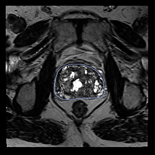
T2 MR of the prostate of one patient before a fusion biopsy, with the gland contoured semi-automatically.
Read before comparing (1)
- Cases. The MR images, the fixed images of ProstateMRUS's pairs, each on its pair's side of the split. The ultrasound images are left out.
| Accuracy | Cost | |||||||
|---|---|---|---|---|---|---|---|---|
| # | Method | Dice | Surface Dice | HD95 mm | Time s | RAM GB | GPU GB | Images |
| 11–2 | KonfAI segmentation app · TotalSegmentator (total_mr), the authors' packageGPUapp | 0.813 ±0.022 | 0.342 ±0.033 | 6.80 ±0.88 | 19 | 11.1 | 2.2 | |
| 21–2 | KonfAI segmentation app · TotalSegmentator (total_mr), KonfAI appGPUapp | 0.803 ±0.026 | 0.344 ±0.040 | 7.23 ±1.20 | 21 | 2.2 | 1.8 | |
The order averages each method's ranks on Dice, Surface Dice, HD95 mm, so the first one is not always the best in every column. ± is the 95% interval of the mean over the 30 cases. Two neighbours are tied when a paired Wilcoxon test on the shared cases does not find the upper one better on Dice (5%): the places a method could hold are then shown under its own. The best value of each column is in colour. Click a row to see its images, a column title to sort by it. Time and memory measured on Workstation.
1 result not ranked: an older run, cases still missing, or a failure on every case
| Accuracy | Cost | ||||||||
|---|---|---|---|---|---|---|---|---|---|
| # | Method | Dice | Surface Dice | HD95 mm | Time s | RAM GB | GPU GB | Machine | Images |
| KonfAI segmentation app · TotalSegmentator (total_mr), KonfAI appGPUappolder run | 0.803 ±0.026 | 0.344 ±0.040 | 7.23 ±1.21 | 4 | 2.0 | 2.0 | Laptop | ||
Dice: how much the organ contours overlap, 1 is perfect. Surface Dice: the share of the contours' surfaces within a tolerance of each other, 1 is perfect. HD95: distance between the contours' surfaces.
Case by case
Click a score to look at that result: the image, the truth in green and the method's map in red. Underlined scores have their image; for the others you see the case without the result.
About the data
- Truth
- contours by a program, verified by people the prostate gland on each image, contoured semi-automatically with Eigen's tools (ProFuse on the MR, the Artemis system on the ultrasound), filled by us on each image's grid
- Source
- www.cancerimagingarchive.net/collection/prostate-mri-us-biopsy
doi.org/10.7937/TCIA.2020.A61IOC1A
doi.org/10.1016/j.juro.2012.08.095
github.com/SlicerHeart/SlicerHeart/blob/master/Docs/ImageImportExport.md#eigen-artemis
zenodo.org/records/10069911 - Licence
- CC BY 4.0
30 cases for tuning, never tested
Prostate-MRI-US-Biopsy-0001/fixed, Prostate-MRI-US-Biopsy-0002/fixed, Prostate-MRI-US-Biopsy-0003/fixed, Prostate-MRI-US-Biopsy-0321/fixed, Prostate-MRI-US-Biopsy-0459/fixed, Prostate-MRI-US-Biopsy-1138/fixed, Prostate-MRI-US-Biopsy-1000/fixed, Prostate-MRI-US-Biopsy-0653/fixed, Prostate-MRI-US-Biopsy-0517/fixed, Prostate-MRI-US-Biopsy-0914/fixed, Prostate-MRI-US-Biopsy-0713/fixed, Prostate-MRI-US-Biopsy-0908/fixed, Prostate-MRI-US-Biopsy-1037/fixed, Prostate-MRI-US-Biopsy-0075/fixed, Prostate-MRI-US-Biopsy-0876/fixed, Prostate-MRI-US-Biopsy-0309/fixed, Prostate-MRI-US-Biopsy-0668/fixed, Prostate-MRI-US-Biopsy-0650/fixed, Prostate-MRI-US-Biopsy-0152/fixed, Prostate-MRI-US-Biopsy-1004/fixed, Prostate-MRI-US-Biopsy-1038/fixed, Prostate-MRI-US-Biopsy-0421/fixed, Prostate-MRI-US-Biopsy-0718/fixed, Prostate-MRI-US-Biopsy-0455/fixed, Prostate-MRI-US-Biopsy-0802/fixed, Prostate-MRI-US-Biopsy-1067/fixed, Prostate-MRI-US-Biopsy-1111/fixed, Prostate-MRI-US-Biopsy-1142/fixed, Prostate-MRI-US-Biopsy-0328/fixed, Prostate-MRI-US-Biopsy-0425/fixed