 fidopt
Pediatric thoracic dynamic MRI
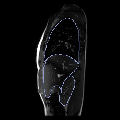
Free-breathing dynamic MRI of one healthy child's thorax and upper abdomen, the end-expiration (fixed) and end-inspiration (moving) volumes of one 4D image, lungs, liver, spleen and kidneys segmented at both (Virtual Growing Child).
Read before comparing (8)
- Pool. Healthy children of the Virtual Growing Child project, University of Pennsylvania and Children's Hospital of Philadelphia. The Dryad abstract speaks of 200 children; its archive, byte for byte the one of the authors' page, holds 141, VGC264 to VGC548, each with both phases and all ten masks.
- Images. Free-breathing 3 T true-FISP. The authors' OFx method picks one sagittal slice per position and phase to build one 4D image over one breathing cycle, then standardises each volume's intensities. A volume stacks slices from different breathing cycles, so steps between neighbouring slices are part of the images. The archive holds only the end-expiration and end-inspiration volumes.
- Pairs. End expiration is fixed and end inspiration moving, as in Learn2Reg's lung CT task. The two share one grid from one acquisition and are used as delivered: the breathing is what is registered. On the test cases, pixels of 1.46 to 1.76 mm in the sagittal plane and 37 to 51 slices 6 mm apart.
- Orientation. The DICOM files hold no position or orientation. prepare.py stacks the slices by location and writes the orientation the anatomy shows, origin at zero, checked on every case from the masks: the right lung and the liver on the right, the lungs above the liver.
- Truth. A pretrained U-Net segmented ten objects at both phases; the authors checked each result by eye and corrected it by hand where needed, under a radiologist with over 25 years of experience in thoracoabdominal MRI. Graded semi-automatic: no agreement is given, and the check let errors through, on VGC548 and VGC352. The 20 cases built first have no such slice.
- Volumes. Masks are drawn on 6 mm slices, so a label gains or loses a whole slab at a time. Between the phases the test cases' liver changes by -9 to +7 %, spleen by -13 to +32 % and kidneys by -31 to +38 %, while the lungs grow by 6 to 28 %. Matching the labels exactly takes changing the small organs' volume, which weigh as much as the lungs in the mean Dice.
- Baseline. Unregistered, the six organs overlap with a mean Dice of 0.84 a pair (0.80 to 0.87; lungs 0.89 and 0.90, liver 0.86, spleen 0.74, kidneys 0.83 and 0.84) and an HD95 of 8.8 mm (6.4 to 12.1).
- Shown apart. Two maps are shown, never ranked. The hemi-diaphragms, sheets one or two pixels thick, score a Dice of 0.05 unregistered, so their HD95, 12.2 mm (8.6 to 17.5), says more. The skin outer boundary barely moves (Dice 0.998, HD95 0.7 mm) and flags a field that tears or shifts the body's outline.
| Accuracy | Smoothness | Cost | ||||||||
|---|---|---|---|---|---|---|---|---|---|---|
| # | Method | Dice | Change | HD95 mm | SDlogJ | Folds % | Time s | RAM GB | GPU GB | Images |
| 1 | DemonsCPUapp | 0.898 ±0.010 | +0.054 | 5.95 ±0.84 | 0.030 | 0.00 | 2 | 0.5 | ||
| 2 | ConvexAdam (MIND)GPUapp | 0.889 ±0.010 | +0.045 | 6.37 ±0.78 | 0.030 | 0.00 | 8 | 2.1 | 0.9 | |
| No registration | 0.844 ±0.012 | 8.80 ±0.93 | 0.000 | 0.00 | ||||||
The order averages each method's ranks on Dice, HD95 mm, SDlogJ, so the first one is not always the best in every column. ± is the 95% interval of the mean over the 14 pairs. Two neighbours are tied when a paired Wilcoxon test on the shared pairs does not find the upper one better on Dice (5%): the places a method could hold are then shown under its own. The best value of each column is in colour. Click a row to see its images, a column title to sort by it. Time and memory measured on Laptop.
Dice: how much the organ contours overlap, 1 is perfect. HD95: distance between the contours' surfaces. SDlogJ: how irregular the deformation is, 0 is perfectly smooth. Folds: share of the image where the deformation folds over itself.
Pair by pair
Click a score to look at that result: the fixed image, and over it the moving image after registration, with the annotations. Underlined scores have their image; for the others you see the pair without the result. A green cell gained against no registration on that pair, a red one lost.
About the data
- Test pairs
- 14 test children (10% of the 141) drawn with seed 2026, then 12 development children (10% of the 127 left): VGC264, VGC265 and VGC266 and the last nine of the seeded order. VGC548 and VGC352, whose labels hold stray slices, are kept out of development. One case a child.
- Truth
- contours by a program, verified by people diaphragm by a program, verified by people body by a program, verified by people the dataset's masks at both phases: left lung (1), right lung (2), liver (3), spleen (4), left kidney (5), right kidney (6)
- Source
- doi.org/10.5061/dryad.vmcvdnczf
www.mipg.upenn.edu/Vnews/mipg_data_share/Thorax_dMRI.html
doi.org/10.1117/12.3006807
doi.org/10.1016/j.media.2021.102088 - Licence
- CC0 1.0
12 pairs for tuning, never tested
VGC264, VGC265, VGC266, VGC484, VGC478, VGC442, VGC338, VGC272, VGC397, VGC322, VGC285, VGC430