 fidopt
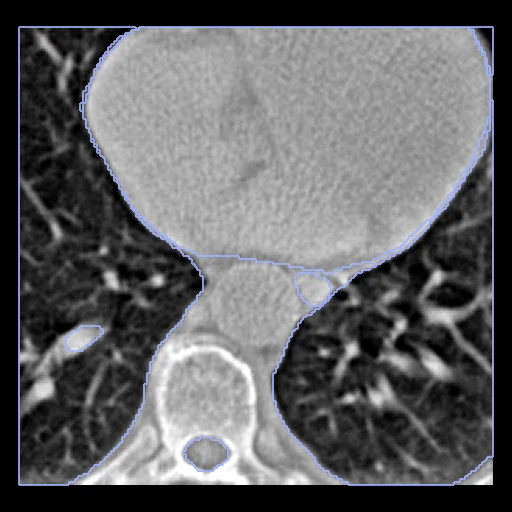
Simulated thoracic CBCT, four clinical contours
Simulated CBCT of the thorax (normalised, no unit) with the clinical contours of the spinal cord, oesophagus, heart and lungs of the planning CT it was simulated from.
Read before comparing (5)
- Images. The deposit's base pair without its geometric augmentations: the CBCT (OS-SART reconstruction) is simulated from the planning CT on the same cropped grid; no real cone-beam acquisition is involved. The CBCT stays on the provider's normalised scale, which differs by patient, without Hounsfield units.
- Truth. LCTSC's clinical contours as the deposit resampled them on the cropped grid, drawn on the planning CT, not on a real CBCT. Voxels two contours claim are in the ignore region.
- Cone. The simulated cone is empty outside its reconstructed field of view; those voxels are in the ignore region.
- Scope. Contours scored on a simulated, normalised CBCT say nothing of real CBCT acquisitions, and a method that expects Hounsfield units has to handle the input itself.
- Overlap. These patients are LCTSC Train patients outside LCTSC's development pairs and outside PsCBCTThorax. A method trained on LCTSC's Train set has seen them and must leave them out. PsCBCTThorax_Seg and PsCBCTThorax_Syn share these patients and this partition; neither shows the other's truth.
| Accuracy | Cost | |||||||
|---|---|---|---|---|---|---|---|---|
| # | Method | Dice | Surface Dice | HD95 mm | Time s | RAM GB | GPU GB | Images |
| 1 | KonfAI segmentation app · IMPACTSeg body, KonfAI appGPUapp1 of 4 structures | 0.809 ±0.024 | 0.612 ±0.088 | 3.08 ±0.69 | 3 | 1.6 | 1.7 | |
The order averages each method's ranks on Dice, Surface Dice, HD95 mm, so the first one is not always the best in every column. ± is the 95% interval of the mean over the 10 cases. Two neighbours are tied when a paired Wilcoxon test on the shared cases does not find the upper one better on Dice (5%): the places a method could hold are then shown under its own. The best value of each column is in colour. Click a row to see its images, a column title to sort by it. Time and memory measured on Workstation.
Dice: how much the organ contours overlap, 1 is perfect. Surface Dice: the share of the contours' surfaces within a tolerance of each other, 1 is perfect. HD95: distance between the contours' surfaces.
Case by case
Click a score to look at that result: the image, the truth in green and the method's map in red. Underlined scores have their image; for the others you see the case without the result.
About the data
- Test cases
- 10 test and 6 development patients, the 16 LCTSC Train patients no other dataset holds: LCTSC uses the 24 provider Test patients and 20 Train patients, PsCBCTThorax the 24 Test patients and 10 of those 20. The test patients were drawn with seed 2026 by site in proportion (2, 4 and 4 from S1, S2 and S3); the other six are the development patients.
- Truth
- contours by experts LCTSC's four clinical contours as the deposit resampled them. Voxels two contours claim, and the empty cone of the simulated CBCT, are in the ignore region rather than given to one structure.
- Source
- zenodo.org/records/5035494
doi.org/10.5281/zenodo.5035494
doi.org/10.1088/1361-6560/abe2eb
www.cancerimagingarchive.net/collection/lctsc
doi.org/10.7937/K9/TCIA.2017.3R3FVZ08 - Licence
- CC BY 4.0 (the Zenodo record); the planning CTs and contours it is made of are LCTSC's, CC BY 3.0
6 cases for tuning, never tested
Train-S1-004, Train-S1-012, Train-S2-006, Train-S2-007, Train-S3-001, Train-S3-012