 fidopt
Multiple sclerosis lesions, Baghdad Teaching Hospital
Brain FLAIR MR of a patient with multiple sclerosis, 2D slices 5 to 8 mm apart at 1.5 T, the lesions outlined by a radiology resident and corrected by two consultants (Muslim et al. 2022).
10 test casesbrainimage MR
Read before comparing (10)
- Cases. The patient's FLAIR and its own mask; the 60 patients were scanned in 20 centres with different parameters. The T1 and T2 are left out: a patient's three sequences share one grid only on Patient-43, and the headers hold no position to bring one onto another. A score here is one from the FLAIR alone.
- Spacing. Every header says 1 x 1 x 1 mm, with no orientation. prepare.py writes on the third axis the slice spacing of the provider's supplementary table 2, 5.2 to 8.3 mm on the test cases. The first two axes keep 1, counting pixels, not millimetres; a pixel is near 0.4 to 1 mm.
- Distances. The surface Dice's 1 mm is one pixel in the slice whatever its true size, and an HD95 in millimetres means nothing across cases. A method that resamples to a physical resolution sees heads up to twice their size.
- Orientation. It differs between cases and is left as delivered. The slices run upwards on most cases and downwards on some; nine test cases are axial and one sagittal (Patient-56), and development holds five axial, four coronal and one sagittal. The patient's left cannot be told, but the one label has no side.
- Truth. A radiology resident outlined the lesions slice by slice on each sequence; a consultant radiologist, then a consultant neurologist, corrected and validated the work. The dataset calls it a consensus, but the readings are not independent, and no agreement between raters is given.
- Values. 64-bit floats holding whole numbers, without a unit, on scales that differ between scanners. The head is not skull-stripped, and no mask is given.
- Room. On the test cases the masks hold 609 to 21,501 voxels of lesion in 13 to 145 pieces, so small lesions weigh on the Dice. Moved by one pixel within the slice, the test masks keep a Dice of 0.74 to 0.94; the surface Dice forgives that move.
- Checks. On the 20 cases built each mask lies on its image's grid, and the lesions sit on hyperintensities of the FLAIR, periventricular and in the white matter.
- Outside numbers. None: the article reports no method, and the dataset belongs to no challenge. The masks are public, so a network trained on the whole archive has seen these patients.
- Overlap. No other dataset of the corpus holds these patients.
No method has a result on every case of this dataset yet.
Case by case
Click a score to look at that result: the image, the truth in green and the method's map in red. Underlined scores have their image; for the others you see the case without the result.
About the data
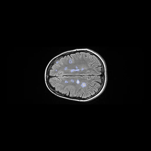
The first test caseClick to look at every case.
- Test cases
- 10 test patients drawn with seed 2026 among the 50 outside development, one FLAIR each; 10 development patients, the last of the same seeded order. The pool is all 60 patients, every mask holding a lesion. No centre is recorded for a patient, so the draw has no strata.
- Truth
- contours by experts the dataset's lesion mask of the FLAIR, drawn slice by slice by a radiology resident with three years of experience, corrected and validated by a consultant radiologist with ten, then by a consultant neurologist of the clinic with eleven
- Source
- doi.org/10.17632/8bctsm8jz7.1
data.mendeley.com/public-api/datasets/8bctsm8jz7
doi.org/10.1016/j.dib.2022.108139 - Licence
- CC BY 4.0
10 cases for tuning, never tested
Patient-14, Patient-17, Patient-53, Patient-58, Patient-23, Patient-12, Patient-54, Patient-32, Patient-27, Patient-22