 fidopt
Colorectal-Liver-Metastases, liver and veins
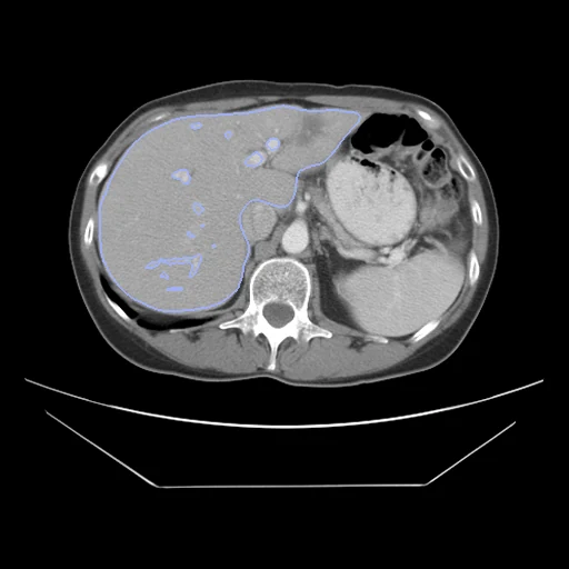
Preoperative portal-venous CT of the liver of two patients with colorectal liver metastases, paired by us, with the liver and its hepatic and portal veins segmented (Colorectal-Liver-Metastases, Memorial Sloan Kettering).
Read before comparing (9)
- Truth. The liver and its hepatic and portal veins, segmented semi-automatically in Scout Liver by one expert radiologist or fellow a scan. The collection says every segmentation was verified, but reports no second reading and no measure of accuracy. The liver label holds the tumours, and the veins are painted over it.
- Veins. How far each venous tree is followed differs from scan to scan: on the 40 scans built, 0.26 to 1.0 of the hepatic veins and 0.34 to 1.0 of the portal veins lie inside the liver. Two patients' trees do not branch alike, so no field overlaps them fully; the vessel overlap measures how far the main trunks come together.
- Left out. The collection's tumour and future liver remnant labels: between two patients neither is one structure seen twice, so their overlap says nothing of a registration.
- Alignment. The scans keep their native voxels, the liver centres 28 to 265 mm apart on the test pairs. The moving image's header carries one translation from elastix mutual information inside each body, table left out, the best of several starts, no annotation used. A rigid fit turns the bodies by 2 to 11 degrees, a posture difference, so only the translation is kept.
- Baseline. With no registration the 10 test pairs start at a Dice of 0.22 (0.11 to 0.31), liver 0.58 (0.32 to 0.77), hepatic veins 0.05 and portal veins 0.02, and an HD95 of 45 mm (26 to 89).
- Scans. The portal-venous phase, 80 s after injection, of a contrast CT made within 6 weeks before the resection, mostly on GE LightSpeed scanners. Each series covers the liver and a few centimetres around it; on 4 of the 40 scans built the liver reaches an end slice and may be cut. Slices lie 1.5 to 7.5 mm apart, and on thick ones the veins are coarse.
- Patients. 197 of 384 consecutive liver resections at Memorial Sloan Kettering, the metastases confirmed by pathology. CRLM-CT-1074, scanned prone, is in neither the test nor the development cases.
- Overlap. CRLMCT_Seg's cases are these pairs' fixed images, each on its pair's side of the split; no other dataset of the corpus holds these scans.
- Outside numbers. None: the pairing is ours.
| Accuracy | Smoothness | Cost | ||||||||
|---|---|---|---|---|---|---|---|---|---|---|
| # | Method | Dice | Change | HD95 mm | SDlogJ | Folds % | Time s | RAM GB | GPU GB | Images |
| 11–2 | DemonsCPUapp | 0.310 ±0.039 | +0.094 | 35.87 ±9.29 | 0.235 | 4.16 | 18 | 2.9 | ||
| 21–2 | ConvexAdam (MIND)GPUapp | 0.281 ±0.063 | +0.065 | 44.93 ±14.60 | 0.127 | 1.50 | 20 | 3.2 | 5.6 | |
| No registration | 0.216 ±0.048 | 45.48 ±13.75 | 0.000 | 0.00 | ||||||
The order averages each method's ranks on Dice, HD95 mm, SDlogJ, so the first one is not always the best in every column. ± is the 95% interval of the mean over the 10 pairs. Two neighbours are tied when a paired Wilcoxon test on the shared pairs does not find the upper one better on Dice (5%): the places a method could hold are then shown under its own. The best value of each column is in colour. Click a row to see its images, a column title to sort by it. Time and memory measured on Laptop.
Dice: how much the organ contours overlap, 1 is perfect. HD95: distance between the contours' surfaces. SDlogJ: how irregular the deformation is, 0 is perfectly smooth. Folds: share of the image where the deformation folds over itself.
Pair by pair
Click a score to look at that result: the fixed image, and over it the moving image after registration, with the annotations. Underlined scores have their image; for the others you see the pair without the result. A green cell gained against no registration on that pair, a red one lost.
About the data
- Test pairs
- 10 test pairs drawn with seed 2026 among the 88 pairs outside development, and 10 development pairs. The 197 patients are put in the seed's order and paired two by two, with no regard to sex, age, scanner or operation; the last, CRLM-CT-1158, is left out.
- Truth
- contours by experts 3 structures, the dataset's segmentations: 1 the liver, its tumours included, then 2 the hepatic veins and 3 the portal veins over it; made semi-automatically in Scout Liver by an expert radiologist or fellow
- Source
- www.cancerimagingarchive.net/collection/colorectal-liver-metastases
doi.org/10.7937/QXK2-QG03
doi.org/10.1038/s41597-024-02981-2 - Licence
- CC BY 4.0
10 pairs for tuning, never tested
1037_1030, 1063_1164, 1054_1176, 1167_1004, 1137_1062, 1003_1091, 1028_1100, 1180_1020, 1134_1013, 1192_1105