 fidopt
Mouse whole-body micro-CT, ten organs
Whole-body micro-CT of one mouse with ten organs segmented: the fixed images of MouseMicroCT's pairs.
10 test casesmouseimage MICROCT
Read before comparing (3)
- Cases. A test pair's fixed image is a test case, a development pair's a development case. The moving images are left out.
- Labels. Native M01 to M05, the OsteoSense group, also hold bone, stomach and part of the thigh muscle; the 15 other native mice hold seven organs (lung, heart, liver, spleen, kidneys, intestine, bladder); the contrast-enhanced maps add the trachea, missing on contrast_M03's last scan.
- Images. The native mice were scanned on a Tomoscope Duo (dual energy, 65 kV), two scans a time point to cover the whole body, the contrast-enhanced mice on an InSyTe (75 kV). The native images are cut around the mouse, each to its own size; the contrast-enhanced ones keep the whole 43 x 34 x 68 mm field. Values are Hounsfield units as delivered (air -1000).
No method has a result on every case of this dataset yet.
Case by case
Click a score to look at that result: the image, the truth in green and the method's map in red. Underlined scores have their image; for the others you see the case without the result.
About the data
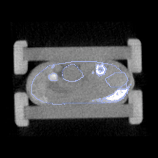
The first test caseClick to look at every case.
- Truth
- contours by a program, verified by people the dataset's organs on each scan, one numbering for all: liver (1), heart (2), lung (3), trachea (4), spleen (5), kidneys (6), stomach (7), intestine (8), bladder (9), bone (10); bone and lung found by a threshold and region growing from a seed point, the heart, liver, kidneys, stomach, intestine and bladder drawn by hand (notes)
- Licence
- CC0 1.0
10 cases for tuning, never tested
native_M10/fixed, contrast_M05/fixed, native_M20/fixed, native_M01/fixed, native_M07/fixed, contrast_M03/fixed, native_M16/fixed, native_M19/fixed, native_M04/fixed, contrast_M08/fixed