 fidopt
Weekly ADC on the MR-Linac, head and neck
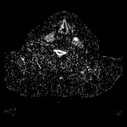
ADC maps of the neck of one head and neck cancer patient on a 1.5 T MR-Linac, before radiotherapy (fixed) and in its third week (moving), from MD Anderson's weekly intra-treatment DWI dataset.
Read before comparing (13)
- Truth. The centres of the dataset's contours, one pair for each structure both sessions hold: the primary tumour, and each node by its name (GTV_N1 to GTV_N4). The ten test pairs hold 20 points, six of them a single one, short of the truth bar's median of ten: ranked apart. The filled contours are shown (dice_tumour), never ranked.
- Shrinking tumours. GTV_P_BL is the tumour before treatment, GTV_P_RD what remains at the week. On the test pairs the residual holds 0.54 to 0.99 of the baseline volume (median 0.80, 9 pairs), the nodes 0.51 to 0.99 (median 0.61, 5 pairs). Uneven shrinkage moves a centre by an unmeasured amount: no method can reach zero.
- Pairs. Node names hold across sessions, and a structure contoured on one session gives no pair. Test cases 1130545713, 1574315175, 1781492641, 2274590586 and 2638457871 are scored on the primary tumour alone, 6394075659 on one node alone.
- Contours. Two radiation oncologists drew them on T2-weighted images that are not released, then carried them to the DWIs by a rigid registration the provider did not measure, which leaves the echo-planar distortion: the truth's own error is unknown. Slices are 4 mm thick, and four test baselines hold a primary tumour under 1 mL (0.4 to 0.8 mL).
- Left out. The weekly sets' GTV_P_BL, the provider's deformable registration of the baseline to each week, and GTV_P_RS, GTV_P_BL minus GTV_P_RD.
- Sessions. The moving image is week 3, else week 2, else week 4; all ten test pairs use week 3, 18 to 38 days after baseline.
- Images. The scanner's ADC maps from echo-planar DWI, b-values 150 and 500 s/mm² only: the stored 16-bit integers without unit, 0 to about 7,200, and 0 in 67 to 79% of a volume's voxels.
- Alignment. Each session has its own frame: as delivered, a structure's two centres lie 9.2 mm apart (2.3 to 35) on the test pairs. The moving header carries one translation from elastix mutual information inside each neck, the best of several starts, no annotation used; a rigid fit turned the sessions 0.2 to 7.6 degrees without moving the centres.
- Baseline. With no registration a case's error, the mean over its points, is 4.3 mm (1.0 to 8.0) on the test cases and the tumour Dice 0.38 (0.05 to 0.66). What remains is the neck bent differently in the treatment mask and the tumours' change.
- Registration. From a former ANTs rigid start, FireANTs with its tutorial settings took the error from 3.1 mm (0.9 to 7.8) to 3.4 mm (1.7 to 7.6), lower on 4 of 10 pairs, though the images agreed better. The anatomy moves and the contour centres do not follow, as HNTSMRG reports: a few centres may not tell a good registration from none.
- Overlap. The patients were treated at MD Anderson Cancer Center (MOMENTUM registry, NCT04075305), as were HNTSMRG's. The anonymised identifiers cannot tell whether a patient is in both; the images differ, T2-weighted there and ADC maps here.
- Outside numbers. No registration result on these images is published.
- No method here does better than no registration on TRE (4.28): as far as these annotations can tell, the pairs already match.
| Accuracy | Smoothness | Cost | ||||||||
|---|---|---|---|---|---|---|---|---|---|---|
| # | Method | TRE mm | Change | rTRE | SDlogJ | Folds % | Time s | RAM GB | GPU GB | Images |
| 1 | DemonsCPUapp | 6.57 ±2.29 | +2.29 | 0.0140 ±0.0041 | 0.053 | 0.04 | 3 | 0.6 | ||
| No registration | 4.28 ±1.73 | 0.0093 ±0.0038 | 0.000 | 0.00 | ||||||
The order averages each method's ranks on TRE mm, SDlogJ, so the first one is not always the best in every column. ± is the 95% interval of the mean over the 10 pairs. Two neighbours are tied when a paired Wilcoxon test on the shared pairs does not find the upper one better on TRE mm (5%): the places a method could hold are then shown under its own. The best value of each column is in colour. Click a row to see its images, a column title to sort by it. Time and memory measured on Laptop.
TRE: distance between matching landmarks. rTRE: the TRE over the length of the image's diagonal, to compare datasets of other sizes and units. SDlogJ: how irregular the deformation is, 0 is perfectly smooth. Folds: share of the image where the deformation folds over itself.
Pair by pair
Click a score to look at that result: the fixed image, and over it the moving image after registration, with the annotations. Underlined scores have their image; for the others you see the pair without the result. A green cell gained against no registration on that pair, a red one lost.
About the data
- Test pairs
- 10 test patients drawn with seed 2026 among the 19 outside development, and 10 development patients, one pair each. The pool is the 29 of the dataset's 30 patients with a structure contoured on both sessions: 2241361130 has none, with no residual primary tumour contoured from the third week on and no node.
- Truth
- landmarks by experts tumour by experts the centre of the primary tumour (GTV_P_BL on the baseline image, the residual GTV_P_RD on the weekly one) and the centre of each involved node by its name (GTV_N1 to GTV_N4), from the dataset's contours, one pair for each structure both sessions hold: 1 to 4 a pair on the test cases, 20 in all
- Licence
- CC BY 4.0
10 pairs for tuning, never tested
5468522794, 2300610798, 7829134765, 1417793418, 2571189939, 2926470717, 2307675387, 1638896444, 2067492015, 3140855008