 fidopt
Human Inner Ear Anatomy
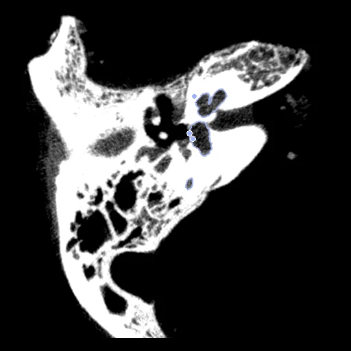
Two cadaver temporal bones scanned by volume CT at 0.0986 mm, with the inner ear's fluid space labelled by hand (Human Inner Ear Anatomy).
Read before comparing (11)
- Specimens. 43 frozen cadaver temporal bones, in-house scans of the paper's Würzburg authors on a Siemens Axiom-Artis DynaCT flat-panel volume CT at 98.6 µm. They are paired two by two in the order of sha256("2026:InnerEarCT:<nn>"), each in one pair.
- Truth. A researcher trained and supervised by an otologic surgeon painted the fluid space by hand in 3D Slicer, helped by thresholds: cochlea, vestibule, and the three semicircular canals as one label. No agreement between raters is given, and the labels say nothing of the bone around. The canals are thin tubes, so their Dice moves with a few voxels.
- Landmarks. The helicotrema, the oval window and the round window, marked with the labels and used at the mm positions 3D Slicer recorded. On the 40 specimens built they lie on their structures within 0.22 mm. Their error is shown, never ranked.
- Sides. The dataset names no side, but the vestibule's side of the three landmarks' plane does: 39 right ears and 4 left (12, 18, 22 and 38). Where a pair joins a right and a left ear, prepare.py mirrors the moving left ear with its labels and landmarks on the same grid: test case 14_38 and development cases 01_22, 06_18 and 29_12.
- Donors. The dataset does not say which specimens share a donor: a mirrored pair may join the two ears of one person.
- Alignment. The two ears come on their own grids, turned 10 to 53 degrees from each other. The moving image's, labels' and landmarks' headers carry one rigid transform from elastix mutual information inside each specimen, no annotation used; both ears keep their voxels. On 41_43, 27_42, 16_39 and 30_17 it lands on the bone around the labyrinth, 2.4 to 5.6 mm from the landmarks.
- Baseline. With no registration the 10 test pairs start at a label Dice of 0.40 (0.04 to 0.62), an HD95 of 2.0 mm (0.7 to 5.6) and a landmark error of 1.9 mm (0.5 to 5.6).
- Rigid only. The three label centres lie within 0.06 to 0.68 mm of one line, so a fit on them is free to turn about it, and three landmarks are too few for an affine fit. The difference in size between two ears is left to the methods.
- Images. 512 x 512 x 405 voxels of 0.0986 mm, the values as delivered in 16 bits; the field is asked on the fixed grid, 106 million voxels. 42.hdf5, the moving image of test pair 27_42, has no header: prepare.py gives it the others' spacing, the identity direction and the origin its landmarks give.
- Overlap. No specimen is in BonyLabyrinth, the corpus's other inner-ear dataset: those are 23 embalmed temporal bones from Bern, scanned on a clinical CT and a micro-CT by other authors.
- Outside numbers. None: the pairing is ours.
| Accuracy | Smoothness | Cost | ||||||||||
|---|---|---|---|---|---|---|---|---|---|---|---|---|
| # | Method | Dice | Change | HD95 mm | TRE mm * | rTRE | SDlogJ | Folds % | Time s | RAM GB | GPU GB | Images |
| 1 | DemonsCPUapp | 0.411 ±0.166 | +0.015 | 2.11 ±0.96 | 2.23 ±1.09 | 0.0272 ±0.0134 | 0.058 | 0.00 | 87 | 13.8 | ||
| No registration | 0.396 ±0.168 | 2.04 ±1.21 | 1.94 ±1.22 | 0.0237 ±0.0149 | 0.000 | 0.00 | ||||||
The order averages each method's ranks on Dice, HD95 mm, SDlogJ, so the first one is not always the best in every column. ± is the 95% interval of the mean over the 10 pairs. Two neighbours are tied when a paired Wilcoxon test on the shared pairs does not find the upper one better on Dice (5%): the places a method could hold are then shown under its own. The best value of each column is in colour. Click a row to see its images, a column title to sort by it. Time and memory measured on Laptop.
* shown, not ranked: on this dataset it is checked against the output of a program.
Dice: how much the organ contours overlap, 1 is perfect. HD95: distance between the contours' surfaces. TRE: distance between matching landmarks. rTRE: the TRE over the length of the image's diagonal, to compare datasets of other sizes and units. SDlogJ: how irregular the deformation is, 0 is perfectly smooth. Folds: share of the image where the deformation folds over itself.
Pair by pair
Click a score to look at that result: the fixed image, and over it the moving image after registration, with the annotations. Underlined scores have their image; for the others you see the pair without the result. A green cell gained against no registration on that pair, a red one lost.
About the data
- Test pairs
- 10 test pairs drawn with seed 2026 among the 11 outside development, and 10 development pairs. The pairs are ours, from the dataset's 43 specimens: specimen 24 is left over and pair 15_32 is in neither set.
- Truth
- contours by experts landmarks by experts the dataset's manual labels of the inner ear's fluid space: 1 cochlea, 2 vestibule, 3 semicircular canals the dataset's three manual landmarks, the helicotrema (apex of the cochlea), the oval window and the round window: their error is shown, never ranked
- Licence
- CC BY 4.0
10 pairs for tuning, never tested
01_22, 29_12, 08_35, 05_19, 20_07, 09_37, 40_03, 34_02, 44_21, 06_18