 fidopt
NeuroPAL atlas (C. elegans heads)
Confocal fluorescence volume of the head of two young-adult C. elegans hermaphrodites, the panneuronal TagRFP-T channel of NeuroPAL, paired by us from the ten worms of the NeuroPAL atlas (DANDI 000715).
Read before comparing (10)
- Images. One channel of a confocal stack, the TagRFP-T that marks every neuron's nucleus with nearly uniform brightness, so all neurons show, named or not. The intestine's granules show at the back of every volume, and a few large round bodies, not annotated, in some heads. Values are 16-bit counts; the brightest nuclei clip at 65535. No mask is given.
- Units. The voxels are 0.21 x 0.21 x 0.75 µm, and the dataset's unit is the µm: the fields and every distance the board gives, the TRE included, are in micrometres.
- Truth. The neuron centres of the DANDI files, paired by name, 184 to 188 pairs a pair, all named by NeuroPAL's designer, Eviatar Yemini; each lies on a nucleus, at most half a voxel off. The original release marks 73 of the 1,903 names as low confidence, 8 to 18 pairs a pair; DANDI drops the mark, and the corpus keeps them.
- Convention. NeuroPAL cannot tell some neurons of a class apart, such as the left from the right AVF, SABV, RIG and RIF. The annotator names them by an educated guess, so 18 to 20 landmark pairs a pair are matched by convention, not identity.
- Swaps. Cell bodies change places within a ganglion from one animal to the next, and neighbours often swap, which no smooth one-to-one deformation can follow: part of what is left on a pair is no registration error, and no method can reach zero.
- Alignment. Each worm keeps its voxels and grid; no transform is computed and no image is resampled. Five worms were imaged from the side and five from above or below, so a pair across the views is a quarter turn apart about the body axis, and the optical axis reads short. Both are left to the methods.
- Baseline. As delivered the three test pairs leave the neurons 13.2 µm apart (7.2 to 16.3), better on every pair than the image centres put together (20.1 µm) or an elastix translation (19.9 µm). 4 moving landmarks of the test pairs lie outside the fixed image's grid.
- Metadata. The NWB files carry metadata copied from another dataset's conversion, such as the strain and the microscope. The corpus reads only the image, the voxel size, the channel indices and the neurons, which follow the original files.
- Scope. Three test pairs separate only large differences between methods. All ten worms are young-adult hermaphrodites of one strain and age, named by one person: the board says nothing of other stages, strains or microscopes, nor of the rest of the body.
- Outside numbers. None: the pairing is ours.
| Accuracy | Smoothness | Cost | ||||||||
|---|---|---|---|---|---|---|---|---|---|---|
| # | Method | TRE µm | Change | rTRE | SDlogJ | Folds % | Time s | RAM GB | GPU GB | Images |
| 1 | ConvexAdam (MIND)GPUapp | 13.02 ±13.92 | −0.19 | 0.0696 ±0.0699 | 0.101 | 0.48 | 7 | 2.5 | 2.2 | |
| No registration | 13.21 ±12.88 | 0.0707 ±0.0641 | 0.000 | 0.00 | ||||||
The order averages each method's ranks on TRE µm, SDlogJ, so the first one is not always the best in every column. ± is the 95% interval of the mean over the 3 pairs. Two neighbours are tied when a paired Wilcoxon test on the shared pairs does not find the upper one better on TRE µm (5%): the places a method could hold are then shown under its own. The best value of each column is in colour. Click a row to see its images, a column title to sort by it. Time and memory measured on Laptop.
TRE: distance between matching landmarks. rTRE: the TRE over the length of the image's diagonal, to compare datasets of other sizes and units. SDlogJ: how irregular the deformation is, 0 is perfectly smooth. Folds: share of the image where the deformation folds over itself.
Pair by pair
Click a score to look at that result: the fixed image, and over it the moving image after registration, with the annotations. Underlined scores have their image; for the others you see the pair without the result. A green cell gained against no registration on that pair, a red one lost.
About the data
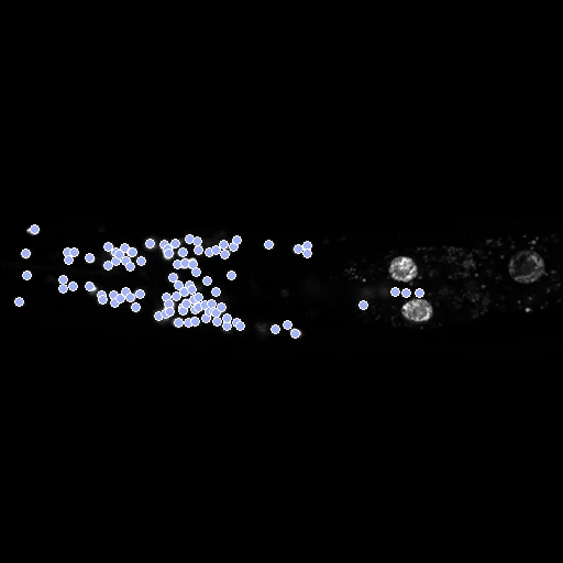
- Test pairs
- The ten worms make five pairs, each worm in one: 3 test pairs and 2 development pairs. Two test pairs join a worm imaged from the side with one imaged from above or below (64 and 20, 38 and 70); one joins two seen from above or below (76 and 56).
- Truth
- landmarks by experts the centres of the neurons named in both worms, 184 to 188 a pair: the names given by Eviatar Yemini (Hobert lab, Columbia University) from each neuron's NeuroPAL colours and position
- Source
- dandiarchive.org/dandiset/000715/0.241009.1514
doi.org/10.48324/dandi.000715/0.241009.1514
zenodo.org/records/3906530
doi.org/10.1016/j.cell.2020.12.012
www.hobertlab.org/wp-content/uploads/2019/06/NeuroPAL-Reference-Manual-v1_small.pdf
pmc.ncbi.nlm.nih.gov/articles/PMC10257892
github.com/focolab/NWBelegans/blob/main/NWB_convert.py - Licence
- CC BY 4.0
2 pairs for tuning, never tested
sub-55-YAaDV_sub-62-YAaLR, sub-7-YAaLR_sub-11-YAaLR