 fidopt
Sunnybrook Cardiac Data
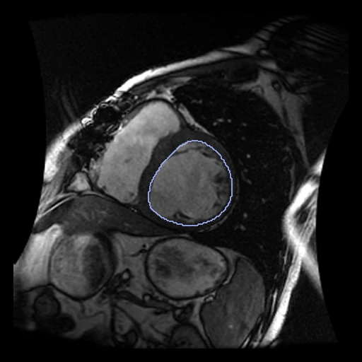
Cine MR of the heart, a short-axis stack of one patient at end diastole (fixed) and end systole (moving) (Sunnybrook Cardiac Data, the MICCAI 2009 left ventricle segmentation challenge).
Read before comparing (8)
- Truth. An experienced cardiologist drew the endocardial contours on every slice of both frames and a second one confirmed them, papillary muscles and trabeculations inside the cavity. prepare.py fills each contour on its slice; the files do not say where a pixel's centre sits, which leaves half a pixel (0.6 to 0.7 mm) of doubt in the plane.
- Labels. One structure, the left ventricular cavity. The files hold the epicardium at end diastole only, on all 45 patients, so the myocardium cannot be scored; the papillary muscle contours are not used.
- Slices. 8 to 16 short-axis slices of 1.37 mm pixels (1.21 mm on SC-HF-NI-36), 8 or 10 mm thick and as far apart (SC-HF-NI-36: 8 mm thick, 10 mm apart). The labels check motion in the plane better than across it: a slice gained or lost weighs a whole slab in the Dice and 8 to 10 mm in the HD95, and nothing between slices is checked.
- Coverage. On 4 of the 10 test cases the end-systolic cavity covers one to three slices fewer than the end-diastolic one, at the base (SC-HF-I-12, SC-HF-NI-33, SC-N-10) or at the apex (SC-HF-NI-33, SC-N-05, SC-N-10).
- Alignment. Cine SSFP at 1.5 T, 20 frames over the cycle, in breath-holds of 10 to 15 s. The two frames of a slice location come from one acquisition on one grid, and the pair is used as delivered. On 5 of the 10 test cases each location has its own acquisition time, so neighbouring slices may be offset by different breath-holds, the same in both frames.
- Frames. The fixed image is the frame contoured with the epicardium, the challenge's end diastole (frame 18 to 20 of 20 on the test cases); the moving image is end systole (frame 5 to 9).
- Baseline. Unregistered, the two cavities overlap with a Dice of 0.67 on average (0.39 to 0.94) and an HD95 of 11.2 mm (3.9 to 17.3). The cavity loses 10 % of its volume on SC-HF-I-09, a dilated ventricle that barely contracts, and 76 % on SC-N-10.
- Source. The CAP's archives hold each patient's study under a new name (SCD0000101 for SC-HF-I-01); prepare.py takes the short-axis cine series the contours number.
No method has a result on every pair of this dataset yet.
| Accuracy | Smoothness | Cost | ||||||||
|---|---|---|---|---|---|---|---|---|---|---|
| # | Method | Dice | Change | HD95 mm | SDlogJ | Folds % | Time s | RAM GB | GPU GB | Images |
| No registration | 0.667 ±0.136 | 11.19 ±3.21 | 0.000 | 0.00 | ||||||
Dice: how much the organ contours overlap, 1 is perfect. HD95: distance between the contours' surfaces. SDlogJ: how irregular the deformation is, 0 is perfectly smooth. Folds: share of the image where the deformation folds over itself.
Pair by pair
Click a score to look at that result: the fixed image, and over it the moving image after registration, with the annotations. Underlined scores have their image; for the others you see the pair without the result. A green cell gained against no registration on that pair, a red one lost.
About the data
- Test pairs
- 10 test pairs drawn with seed 2026 among the 35 outside development, two or three from each pathology group in proportion: heart failure with and without infarction, hypertrophy and healthy, 12, 12, 12 and 9 patients. 10 development pairs.
- Truth
- contours by experts the left ventricular cavity inside the endocardial contours a cardiologist drew on each slice of the two frames
- Licence
- CC0 1.0 (the authors ask that Radau et al., MIDAS Journal 2009, be cited)
10 pairs for tuning, never tested
SC-HF-I-01, SC-HF-I-02, SC-HF-I-04, SC-HF-NI-31, SC-HYP-09, SC-HF-NI-12, SC-HYP-06, SC-HF-I-05, SC-HF-NI-13, SC-HYP-08