 fidopt
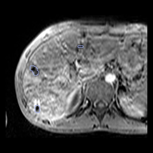
Liver lesions, curated MRI subset
One liver MR scan, most often a contrast-enhanced T1, with a manual liver-lesion mask, from the MR part of the mixed-modality archive released for TotalSegmentator liver_lesions_mr.
Read before comparing (6)
- Pool. The archive mixes CT and MR with no modality metadata. Of its 750 scans, 13 have direction cosines ITK cannot read and 29 an empty mask; of the 708 left, 409 are CT by their air values and 299 are not. An axial slice through each of the 299 shows a liver MR with the mask on a lesion; confirmed_mri.json pins their SHA256.
- Images. One MR channel a case, non-negative 32-bit floats, mostly contrast-enhanced T1 with some coronal acquisitions. Intensities, spacing, origin, direction and field of view are as delivered, with no registration or normalisation.
- Truth. Manual masks, drawn slice by slice on the axial slices. On some cases the drawn extent jumps from one slice to the next, striping the coronal and sagittal planes; on test case 7yhp8iiNtcaBGEiZ (219 mL of lesions) whole rows differ between neighbouring slices. The truth is kept as delivered, and the surface Dice weighs such borders.
- Positives only. Every case holds a lesion: the cases do not measure specificity on lesion-free MR.
- Seen. The archive is the training set of TotalSegmentator liver_lesions_mr: that model, or features derived from it, has seen this truth and cannot claim an independent test result.
- Overlap. Links to patients of other datasets are unavailable. No two of the 299 share a voxel grid, and none has the grid of one of the corpus's 9,968 other MR images: that rules out exact or cropped copies, not a patient scanned twice or an image resampled.
No method has a result on every case of this dataset yet.
Case by case
Click a score to look at that result: the image, the truth in green and the method's map in red. Underlined scores have their image; for the others you see the case without the result.
About the data
- Test cases
- 29 test scans drawn with seed 2026, then 27 development scans, the last of the same seeded order. The pool, confirmed_mri.json, is the archive's 299 MR scans with a non-empty mask on their image's grid, out of 750. The identifiers are random: one patient may have several scans here, which the corpus cannot tell.
- Truth
- contours by experts Manual liver-lesion segmentations as described by the Zenodo release; all nonzero voxels belong to the single liver_lesion class. The record does not describe lesion subtypes or independent multi-rater adjudication.
- Source
- zenodo.org/records/20272348
doi.org/10.5281/zenodo.20272347
github.com/wasserth/TotalSegmentator - Licence
- CC BY 4.0 (Zenodo record 20272348). The nnU-Net dataset.json template inside the archive says Apache 2.0; the record licence is followed.
27 cases for tuning, never tested
dlTGgKSDmODwmecI, ko2BfRinHpGls3Cj, md8DZvWccfR4DzT2, 0BJc4QeljgijtV18, 6qdayzWvUg9JGPoi, Oqbw7bnOPuZz0Kp3, VFNH9RGXiG3tttIK, jXyqes8FXo4vbnBA, 60R1n5m3oTXaLAT8, CCFeOrz2kLk39xO2, 3rjV2LOgfz73VfyP, V1gID29R4S6gjg07, Jnyjt1vO7qngUpZ2, rFE45S86E0VTyjtB, nHCKwIxXbK4zrWD9, WzAgG0nX0AbXZFd6, qNTEKAnj2SbtBl7d, XA0MGLipKLqa0aIZ, eLxzK6lFjhQVlKyO, AkIqZyYgeG1IrrPN, gtVuZNIBhS7dJtOi, qNsbTqlFkrF0Yv3J, xxCsOE8tjGQllAsE, shgniT1q0YodO2lD, 0x9VWUFUAedaos47, cg05sKi8bljvzXLV, rzW9iOZ5E1UdTFxl