 fidopt
4D-Lung, tumour and lymph nodes on 4D CT
One respiratory phase of a 4D fan-beam CT of a patient treated by chemoradiotherapy for locally advanced non-small cell lung cancer, with the tumour and involved lymph nodes contoured on it (TCIA 4D-Lung).
10 test caseslungimage CT
Read before comparing (8)
- Cohort. 20 patients treated at one centre by concurrent chemoradiotherapy for stage IIA to IIIB non-small cell lung cancer. Each has a planning 4D CT on a Philips Brilliance Big Bore, 3 mm slices, ten phases from 0%, the end of inhalation, to 90%, repeated about weekly for most. The 4D cone-beam CTs carry no contour.
- Truth. Several physicians drew the tumour and visible involved nodes by hand on one phase, copied them to the nine others by rigid registration and adjusted them on each; one experienced radiation oncologist reviewed every structure.
- Nodes. Patients 104 and 106 have no node structure: on their four cases, two of them test cases, a method that writes nodes scores 0 for them. The paper does not say whether they have no involved node or none was drawn.
- Not labelled. The lungs, heart, oesophagus and spinal cord, which are on almost no image but the 0% phase of seven patients' planning CT (LCTSC_Seg has them); a vertebra and the carina, drawn as marks; the gold fiducials of patients 103, 104 and 106, which stay visible in or near the tumour.
- Cases. Two cases a patient, so the ten test cases are five patients: the 50% phase of the first and last contoured 4D CT, 27 to 64 days apart, the tumour smaller on the second for 112, 116 and 119; or, for 103, 104 and 106 with one contoured 4D CT, its 0% and 50% phases. Patient 113's spinal hardware streaks its images.
- Artefacts. The paper warns that 4D CT sorting artefacts are common, steps or doubled edges at the diaphragm and sometimes at the tumour; the contours follow the image as it is.
- Checks. Each tumour contour holds a mean of -78 to 47 HU inside and -518 to -69 HU in the 3 pixels around it on 19 cases; on development case 115_2 its 4 ml are mostly aerated lung, a tumour nearly gone. Patient 118's structure sets name another phase's series as their reference, but the contours fit the 50% image they are laid on.
- Overlap. L2R_ThoraxCBCT holds ten of the 20 patients, matched by rigid registration of its cone-beam CTs (correlation 0.80 to 0.996 with one patient, under 0.72 with every other): they are left out. Lung4D_CBCTCT_Syn uses the other ten in the same partitions, so a phase given here as input is a CT that dataset hides as its target. Both datasets are kept knowing this.
No method has a result on every case of this dataset yet.
Case by case
Click a score to look at that result: the image, the truth in green and the method's map in red. Underlined scores have their image; for the others you see the case without the result.
About the data
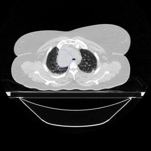
The first test caseClick to look at every case.
- Test cases
- 10 test and 10 development cases, two phases a patient, from the 10 patients L2R_ThoraxCBCT does not hold. In the order of sha256("2026:4D-Lung:<patient>") the first five patients give the test cases and the last five the development cases.
- Truth
- contours by experts the primary tumour (1, the provider's Tumor) and the involved lymph nodes (2, its LN and LN2) drawn by hand on this phase for the study and reviewed by one radiation oncologist
- Source
- www.cancerimagingarchive.net/collection/4d-lung
doi.org/10.7937/K9/TCIA.2016.ELN8YGLE
doi.org/10.1002/mp.12059 - Licence
- CC BY 3.0
10 cases for tuning, never tested
104_1, 107_1, 118_2, 104_2, 115_2, 103_2, 118_1, 107_2, 103_1, 115_1