 fidopt
BUS-BRA, breast ultrasound tumours outlined by hand
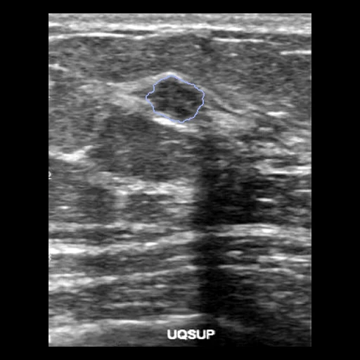
A 2D B-mode breast ultrasound image of a woman's biopsy-proven tumour, benign or malignant, outlined by hand by a senior ultrasonographer: one image a patient of BUS-BRA's 1,064 (National Cancer Institute, Rio de Janeiro).
Read before comparing (8)
- Images. 1,875 grey 8-bit PNGs of 1,064 women examined at the National Cancer Institute in Rio de Janeiro on four scanners (GE Logiq 5 and 7, Toshiba Aplio 300, U-Systems), 240 to 579 pixels wide. No pixel size is given, so distances are in pixels.
- Marks. Calipers and the scanner's text (breast side, quadrant) are burnt into many images, kept as delivered.
- Patients. Each woman has one tumour, in one image (253 women) or two (811, the provider's left and right views, IDs -l and -r). Only the first is used, so a tumour's two views are never on two sides. The test cases hold 20 benign and 10 malignant tumours.
- Truth. One ultrasonographer outlined every tumour; the paper gives no second reading and no agreement figure. A mask holds the tumour only and is never empty.
- Tolerance. 2 pixels. On the test images the truth grown or thinned by one pixel keeps a Dice of 0.98 and a surface Dice of 0.995 to 0.997 at 1 pixel, 0.999 to 1.0 at 2.
- Start. On the 30 test images the whole image scores a Dice of 0.16 and its dark pixels (under Otsu's threshold) 0.19, with a surface Dice of 0.05 at 2 pixels: a tumour covers 10% of its image on average (1 to 49%).
- Overlap. No other dataset of the corpus holds breast ultrasound. In SonoCorpus's per-file MD5 manifest (Zenodo 22770825) no BUS-BRA image is identical, as stored or once converted, to an image of its 52 other sets, BUSI, BUSI-WHU and BrEaST among them. The check finds the known copies inside BUSI, DDTI and TN-SCUI, but cannot see a copy re-encoded or cropped.
- Outside numbers. SonoBase was evaluated on BUS-BRA, not trained on it.
No method has a result on every case of this dataset yet.
Case by case
Click a score to look at that result: the image, the truth in green and the method's map in red. Underlined scores have their image; for the others you see the case without the result.
About the data
- Test cases
- 30 test patients drawn with seed 2026 among the 1,064, and 30 development patients, the last of the same seeded order. One image a patient, its first view. The provider gives cross-validation folds but no test split.
- Truth
- contours by experts the tumour outlined by hand on every image by the senior ultrasonographer who also gave its BI-RADS category (Gómez-Flores et al. 2024); the pathology, 722 benign and 342 malignant patients, proven by biopsy
- Source
- zenodo.org/records/8231412
doi.org/10.5281/zenodo.8231412
doi.org/10.1002/mp.16812
pubmed.ncbi.nlm.nih.gov/37937827
github.com/wgomezf/BUS-BRA - Licence
- CC BY 4.0 (the Zenodo record), with an MIT-style permission notice in the archive's LICENSE.txt; the paper must be cited
30 cases for tuning, never tested
bus_0045-l, bus_0999-l, bus_0842-s, bus_0365-l, bus_0381-l, bus_0031-s, bus_0030-s, bus_1016-l, bus_0532-l, bus_0482-l, bus_0434-l, bus_0167-l, bus_0441-l, bus_0216-l, bus_0408-l, bus_0427-l, bus_0909-l, bus_0632-l, bus_0423-l, bus_0422-l, bus_0007-l, bus_0176-l, bus_1037-l, bus_0377-l, bus_0517-l, bus_0062-l, bus_0264-l, bus_0491-l, bus_0462-l, bus_0559-l