 fidopt
Vestibular-Schwannoma-SEG
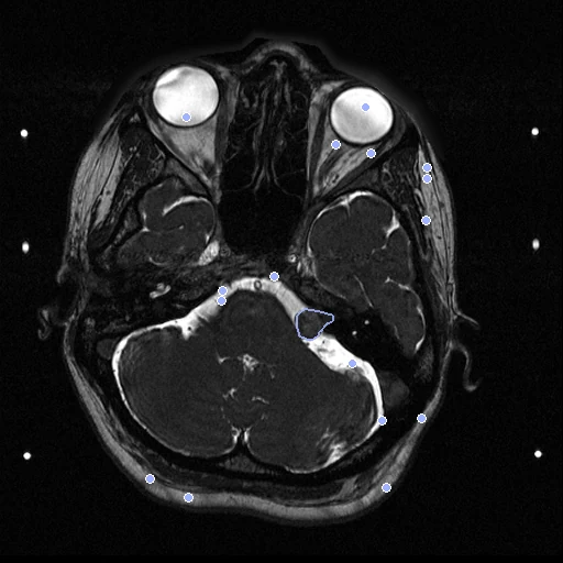
High-resolution T2 (fixed) and contrast-enhanced T1 (moving) MR of the head of one patient with a vestibular schwannoma, from one Gamma Knife session in a stereotactic frame, the T1 deformed by a known warp (Vestibular-Schwannoma-SEG).
Read before comparing (7)
- Truth. As delivered the two series are within 0.4 mm of the frame transform (1.4 mm at most) and leave nothing to register, so the corpus deforms the T1 by a known cubic B-spline (knots 40 mm apart, coefficients within 15 mm, datasets/VestibularSchwannoma/warp/). The labels are warped with the image, and 300 points drawn in the fixed image's head are paired through the transform, every pair inside both images.
- Labels. The treating neurosurgeon and physicist contoured the tumour and the cochlea once, in consensus, and Leksell GammaPlan exported them into the other image through the transform it computed from the frame's fiducials. Label 1 is the case's first structure, the tumour; later volumes and *Skull are left out.
- Label margin. Each label is its image's RT structure set, a contour drawn on the other image interpolated onto this one's slices. Through the frame transform the two maps overlap at a Dice of 0.84 on the 12 test pairs (tumour 0.93, cochlea 0.74), against 0.79 as delivered: label scores move within that margin, while the landmarks are exact.
- Frame error. The provider does not state the fiducial registration's accuracy, and one affine transform cannot hold a difference of distortion between the sequences: errors are measured against that transform, not the anatomy, and its residual, 0.4 mm on average, stays in the truth.
- Alignment. As delivered the two series share the scanner's frame with the head fixed, 0.06 to 1.42 mm from the frame transform at the contour points (0.37 mm on average); each image keeps its own grid and the field is asked on the T2's. With the known warp the 12 test pairs start at a TRE of 4.70 mm (4.26 to 5.03) and a label Dice of 0.25 (0.00 to 0.55), tumour 0.45 and cochlea 0.06.
- Scope. The board measures how well a method recovers a synthetic smooth deformation between two MR contrasts of one head, not a real one; its labels are small targets, the tumour and the cochlea.
- Field of view. The T2's field lies inside the T1's but for up to 7 mm at the edge of the slice and 2 mm below. Both images show the indicator box's fiducials at the edges of the slices.
| Accuracy | Smoothness | Cost | ||||||||||
|---|---|---|---|---|---|---|---|---|---|---|---|---|
| # | Method | Dice | Change | HD95 mm | TRE mm | rTRE | SDlogJ | Folds % | Time s | RAM GB | GPU GB | Images |
| 1 | ConvexAdam (MIND)GPUapp | 0.700 ±0.056 | +0.445 | 1.65 ±0.30 | 1.71 ±0.22 | 0.0054 ±0.0007 | 0.072 | 0.04 | 8 | 2.8 | 3.9 | |
| 2 | NiftyRegCPUapp | 0.565 ±0.083 | +0.310 | 2.51 ±0.49 | 1.90 ±0.14 | 0.0060 ±0.0005 | 0.081 | 0.32 | 62 | 1.4 | ||
| No registration | 0.255 ±0.089 | 5.01 ±0.98 | 4.70 ±0.14 | 0.0149 ±0.0004 | 0.000 | 0.00 | ||||||
The order averages each method's ranks on Dice, HD95 mm, TRE mm, SDlogJ, so the first one is not always the best in every column. ± is the 95% interval of the mean over the 12 pairs. Two neighbours are tied when a paired Wilcoxon test on the shared pairs does not find the upper one better on Dice (5%): the places a method could hold are then shown under its own. The best value of each column is in colour. Click a row to see its images, a column title to sort by it. Time and memory measured on Laptop.
Dice: how much the organ contours overlap, 1 is perfect. HD95: distance between the contours' surfaces. TRE: distance between matching landmarks. rTRE: the TRE over the length of the image's diagonal, to compare datasets of other sizes and units. SDlogJ: how irregular the deformation is, 0 is perfectly smooth. Folds: share of the image where the deformation folds over itself.
Pair by pair
Click a score to look at that result: the fixed image, and over it the moving image after registration, with the annotations. Underlined scores have their image; for the others you see the pair without the result. A green cell gained against no registration on that pair, a red one lost.
About the data
- Test pairs
- 12 test pairs drawn with seed 2026 from a pool of 124 patients, then 11 development pairs. Of the 242 patients, the pool keeps the 140 with both series on NBIA and a contour named cochlea, so every case is scored at two places, less the 15 of VestibularSchwannoma_T1T2_Syn, so no image is in two datasets, and VS-SEG-026, once one of its development patients.
- Truth
- contours a known deformation landmarks a known deformation the tumour and the cochlea, drawn once by the treating neurosurgeon and physicist and exported into each image by the planning software, then carried through the known deformation 300 points drawn inside the head of the fixed image, paired through the known deformation
- Source
- www.cancerimagingarchive.net/collection/vestibular-schwannoma-seg
doi.org/10.7937/TCIA.9YTJ-5Q73
doi.org/10.1038/s41597-021-01064-w
github.com/KCL-BMEIS/VS_Seg - Licence
- CC BY 4.0
11 pairs for tuning, never tested
VS-SEG-001, VS-SEG-002, VS-SEG-003, VS-SEG-230, VS-SEG-019, VS-SEG-149, VS-SEG-045, VS-SEG-050, VS-SEG-165, VS-SEG-093, VS-SEG-071