 fidopt
FDG-PET-CT-Lesions (autoPET)
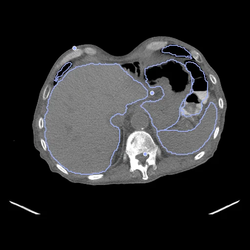
Whole-body CT (fixed) and FDG-PET (moving) of one PET/CT session of a patient with melanoma, lymphoma or lung cancer or of a negative control, the PET deformed by a known smooth transform (autoPET).
Read before comparing (12)
- Alignment. The CT and then the PET of one session on one Siemens Biograph mCT, in one frame of reference: the scanner aligns the pair. The corpus deforms the PET by a known cubic B-spline, knots 40 mm apart and coefficients within 15 mm, and the truth follows it exactly.
- Truth. 300 points drawn in the CT's foreground, above its Otsu threshold, are paired through the transform, each pair inside both images. One label map on the CT's grid goes to the CT as it is and to the PET through the deformation, so the labels score the deformation, not the agreement of two readers.
- Labels. TotalSegmentator's 24 organ classes, run by us on the CT and numbered as it numbers them, under the lesions as label 100, whose score is a case's lesion Dice. Its 93 other classes are left out: the points sample the deformation there. The adrenal glands and kidney cysts are small, and their Dice moves the most.
- Lesions. All FDG-avid tumour lesions, drawn slice by slice on the PET by an experienced radiologist with the CT beside it, after the clinical reading agreed on them. A negative study holds no lesion; its organs still give it a label score. Test case PETCT_41fadf6520_2003-02-27 has one lesion of 31 PET voxels.
- Motion. The scanner's alignment is not perfect near the diaphragm, in the bowel and at the bladder: the PET averages 1 to 4.8 minutes a bed position and starts 3 to 15 minutes after the CT, while the bladder fills. The truth follows the scanner's frame, so a method that matches the PET's own anatomy there is scored off, the same for every method.
- Images. The PET is resampled linearly onto the CT's grid by the deformation and cut to its field of view: 0.74 to 0.96 mm in plane, slices 1 or 2.5 mm apart on the test pairs. PET values are Bq/mL; the files give no SUV.
- Defacing. The public images are the collection's defaced version 2: from the base of the neck up, each image has a box blanked separately, their lower edges 1 to 52 mm apart on the 12 cases measured. Near the neck one image can show anatomy where the other is blank.
- Points. On the ten test pairs drawn first, 89 of the 3,000 points lie where the PET shows almost no activity, mostly on the table and in neck strips blanked on the PET only; they score only how a method extends its field.
- Baseline. With no registration the 30 test pairs score a TRE of 4.61 mm (4.38 to 4.94) and an organ Dice of 0.77 (0.70 to 0.82).
- Methods. The PET shows tracer uptake, not anatomy: a method driven by intensities tends to pull uptake onto the CT's structures rather than undo the known deformation, and the deformable baselines run so far, IMPACT-Reg and ConvexAdam included, do worse than no registration. A method has to read the anatomy the PET barely shows, or keep its deformation small where uptake is.
- Pool. 1,014 studies of 900 patients from University Hospital Tübingen: 188 melanoma, 168 lung cancer, 145 lymphoma and 513 negative by the clinical file. The first development studies were the first, alphabetically, of the melanoma and lung cancer groups, so that tuning sees lesions; the lymphoma group's, PETCT_0223010e46_2003-09-04, was swapped out, so no development study has lymphoma. The others are the last of the seeded order.
- Outside numbers. None: the pair is the scanner's and the deformation ours. autoPET is a segmentation challenge on these images.
No method has a result on every pair of this dataset yet.
| Accuracy | Smoothness | Cost | ||||||||||
|---|---|---|---|---|---|---|---|---|---|---|---|---|
| # | Method | Dice | Change | HD95 mm | TRE mm | rTRE | SDlogJ | Folds % | Time s | RAM GB | GPU GB | Images |
| No registration | 0.769 ±0.011 | 4.64 ±0.08 | 4.61 ±0.05 | 0.0039 ±0.0002 | 0.000 | 0.00 | ||||||
1 result not ranked: an older run, pairs still missing, or a failure on every pair
| Accuracy | Smoothness | Cost | |||||||||||
|---|---|---|---|---|---|---|---|---|---|---|---|---|---|
| # | Method | Dice | Change | HD95 mm | TRE mm | rTRE | SDlogJ | Folds % | Time s | RAM GB | GPU GB | Machine | Images |
| ConvexAdam (MIND)GPUapp7/30 pairs | 0.707 ±0.033 | −0.062 | 8.21 ±1.51 | 9.36 ±0.65 | 0.0090 ±0.0007 | 0.178 | 6.02 | 61 | 7.2 | 22.3 | Laptop | ||
Dice: how much the organ contours overlap, 1 is perfect. HD95: distance between the contours' surfaces. TRE: distance between matching landmarks. rTRE: the TRE over the length of the image's diagonal, to compare datasets of other sizes and units. SDlogJ: how irregular the deformation is, 0 is perfectly smooth. Folds: share of the image where the deformation folds over itself.
Pair by pair
Click a score to look at that result: the fixed image, and over it the moving image after registration, with the annotations. Underlined scores have their image; for the others you see the pair without the result. A green cell gained against no registration on that pair, a red one lost.
About the data
- Test pairs
- 30 test studies drawn by diagnosis with seed 2026, then 30 development studies, one study a patient. The pool is 860 of the 900 patients, once the patients of AutoPET_CTPET_Syn and AutoPET_Seg are left out. Development study PETCT_0223010e46_2003-09-04 was swapped for the next study of the seeded order: its CT of 2,651 slices makes the known-warp build exceed the build's memory cap.
- Truth
- contours a known deformation landmarks a known deformation one map on the CT's grid given to both images, the lesions drawn on the PET by the dataset's reader (label 100) over TotalSegmentator's 24 organ classes run by us on the CT, then carried through the known deformation 300 points drawn in the CT's foreground, paired through the known deformation
- Source
- www.cancerimagingarchive.net/collection/fdg-pet-ct-lesions
doi.org/10.7937/gkr0-xv29
doi.org/10.1038/s41597-022-01718-3 - Licence
- CC BY 4.0
30 pairs for tuning, never tested
PETCT_0011f3deaf_2003-03-23, PETCT_0117d7f11f_2001-09-13, PETCT_64aff75516_2006-01-20, PETCT_30e2b83b74_2002-09-01, PETCT_5420205a5f_2006-12-07, PETCT_9b1bb2165a_2002-10-13, PETCT_6f46454a8d_2005-09-03, PETCT_2f587d00b1_2004-09-27, PETCT_2791a0b3c2_2005-08-29, PETCT_83e4c5b578_2006-03-11, PETCT_9a2bfe901f_2005-08-06, PETCT_71ae114917_2003-04-13, PETCT_e9feb1135e_2005-07-01, PETCT_515c40c4a6_2006-07-24, PETCT_eee2ede9ad_2006-11-24, PETCT_ed7abf4433_2002-12-06, PETCT_7abe274c10_2006-09-21, PETCT_1a8bd52f71_2006-05-05, PETCT_e68c4577d8_2007-05-25, PETCT_06d55e8295_2003-04-17, PETCT_a4ccc397a9_2002-11-30, PETCT_e207c7a1a4_2005-12-23, PETCT_b1d6679284_2007-05-28, PETCT_c1c9e78e0e_2007-05-14, PETCT_bd7603e9df_2005-05-06, PETCT_87afc291ec_2003-07-20, PETCT_72d2d6de52_2005-08-05, PETCT_d951eeb735_2003-01-23, PETCT_1f65acff65_2007-05-06, PETCT_274688d0a8_2007-03-24