 fidopt
Human Bony Labyrinth
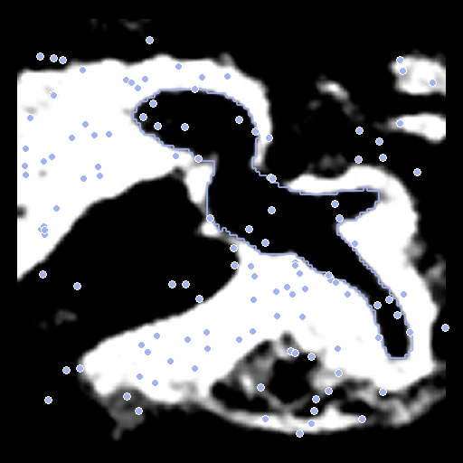
Clinical CT (fixed) and micro-CT (moving) of one human temporal bone specimen, cropped around the inner ear, the micro-CT deformed by a known smooth transform (Human Bony Labyrinth).
Read before comparing (9)
- Alignment. The provider registered each micro-CT rigidly to its CT (normalized mutual information in Amira) and delivers both in one frame: the two expert labels overlap with a Dice of 0.94 (0.88 to 0.96) on the test cases, their centroids 0.03 to 0.22 mm apart. That leaves nothing to register, and the corpus adds no start of its own.
- Warp. The corpus deforms the micro-CT by a known cubic B-spline drawn from the case's seed, knots 4 mm apart and coefficients within 1.5 mm: VestibularSchwannoma's warp scaled by a tenth, since the crops are 11 to 23 mm a side. The labels and points follow it exactly. The deformation is ours, not the body's: the bony labyrinth is rigid.
- Baseline. With no registration the ten test cases score a TRE of 0.46 mm (0.39 to 0.50) and 0.79 mm at the 95th percentile, two to five CT voxels, and a label Dice of 0.78 (0.73 to 0.82): the board measures the last half millimetre.
- Labels. The bony labyrinth (cochlea, vestibule and semicircular canals), labelled once in each image by an expert with region growing, thresholding and manual correction, and reviewed by an experienced otologist. Two readings at two resolutions overlap at a Dice of 0.94, the most a perfect registration can score here; the landmarks are exact.
- Landmarks. Five per specimen, placed by an experienced otologist in the frame both images share: the centres of the round window, the cochlear basal turn, the oval window and the vestibule, and the helicotrema. 300 more points drawn in the CT's bone (Otsu) score the rest of the crop; a point the warp puts outside the micro-CT is drawn again.
- Images. The micro-CT (0.061 mm isotropic) is resampled linearly onto the CT's grid (0.156 x 0.156 x 0.2 mm) through the warp, without smoothing, so it loses detail and aliases some noise. It is not in Hounsfield units, reaching 15673 where the CT stops at 3071, and is given as 'microct' so that no CT window applies. Several micro-CTs hold a small unexplained object at 15673.
- Pool. 23 specimens from 23 donors (Institute of Anatomy, University of Bern), 15 embalmed with formalin (F01 to F15) and 8 with Thiel (T01 to T08).
- Scope. The board measures how well a method recovers a synthetic deformation across a change of modality and resolution on a small rigid structure, not real deformations. The CT to CT pairs between specimens are not built.
- No method here does better than no registration on TRE (0.46): as far as these annotations can tell, the pairs already match.
| Accuracy | Smoothness | Cost | ||||||||||
|---|---|---|---|---|---|---|---|---|---|---|---|---|
| # | Method | Dice | Change | HD95 mm | TRE mm | rTRE | SDlogJ | Folds % | Time s | RAM GB | GPU GB | Images |
| 1 | ConvexAdam (MIND)GPUapp | 0.927 ±0.006 | +0.148 | 0.17 ±0.02 | 0.46 ±0.10 | 0.0149 ±0.0029 | 0.123 | 1.85 | 6 | 2.1 | 0.6 | |
| No registration | 0.780 ±0.021 | 0.48 ±0.05 | 0.46 ±0.02 | 0.0150 ±0.0010 | 0.000 | 0.00 | ||||||
The order averages each method's ranks on Dice, HD95 mm, TRE mm, SDlogJ, so the first one is not always the best in every column. ± is the 95% interval of the mean over the 10 pairs. Two neighbours are tied when a paired Wilcoxon test on the shared pairs does not find the upper one better on Dice (5%): the places a method could hold are then shown under its own. The best value of each column is in colour. Click a row to see its images, a column title to sort by it. Time and memory measured on Laptop.
Dice: how much the organ contours overlap, 1 is perfect. HD95: distance between the contours' surfaces. TRE: distance between matching landmarks. rTRE: the TRE over the length of the image's diagonal, to compare datasets of other sizes and units. SDlogJ: how irregular the deformation is, 0 is perfectly smooth. Folds: share of the image where the deformation folds over itself.
Pair by pair
Click a score to look at that result: the fixed image, and over it the moving image after registration, with the annotations. Underlined scores have their image; for the others you see the pair without the result. A green cell gained against no registration on that pair, a red one lost.
About the data
- Test pairs
- 10 test specimens drawn with seed 2026 among the 13 outside development, stratified by fixation: 7 formalin and 3 Thiel. 10 development specimens. F12 stays in the pool but is neither a test nor a development case: in the provider's F12.zip its CT label file is byte for byte its CT.
- Truth
- contours a known deformation landmarks a known deformation the bony labyrinth, labelled by an expert in the CT and, separately, in the micro-CT, then carried through the known deformation the dataset's five anatomical landmarks and 300 points drawn in the CT, paired through the known deformation
- Source
- zenodo.org/records/3355272
doi.org/10.1016/j.dib.2019.104782
doi.org/10.1007/978-3-030-32254-0_1 - Licence
- CC BY 4.0
10 pairs for tuning, never tested
F01, F02, T01, T04, F05, T07, F11, F13, T05, F15