 fidopt
MR enterography bowel segments
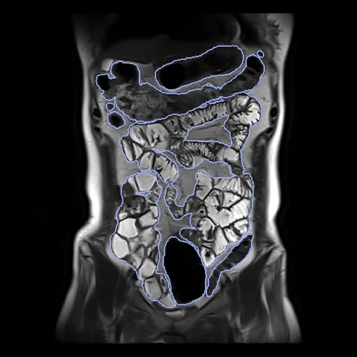
Coronal T2-weighted MR enterography at 3 T of one patient with Crohn's disease, the bowel drawn in ten segments from the stomach to the rectum (Zhong et al., Sci Data 2025).
Read before comparing (8)
- Scans. One coronal HASTE breath-hold on a Siemens Magnetom Prisma or Vida, the bowel filled with mannitol and peristalsis held back by raceanisodamine. The files do not name the scanner, so the strata follow the pixel size: 1.41 mm for the Prisma, 1.17 mm for the Vida. Case 5, at 1.04 mm, matches neither and counts with the Prisma.
- Images. Every file puts its slices 4.8 mm apart where the paper gives sections 4 mm thick; the files' spacing is kept. Values are the files' own 16-bit integers, without a unit. No mask is given.
- Orientation. The files carry no orientation. prepare.py sets the one the anatomy shows on all 114 label maps, a rotation and no mirror: columns from right to left, rows from head to feet, slices from front to back. Liver, spleen and kidneys on test cases 37 and 40, and TotalSegmentator on case 37, confirm it.
- Truth. A pre-trained model's draft, later a network trained on the first batch, refined slice by slice by two radiologists of 3 and 6 years' experience and reviewed to consensus by an abdominal radiologist of more than 10 years. No agreement between readers is given. Label 3 holds the jejunum and ileum together, whose boundary MR often does not show.
- Missing segments. 51 of the 114 maps hold no appendix and case 106 no cecum; a case is scored on the segments its map holds. On the test cases the appendix is drawn on 5 (37, 60, 61, 73, 90), at 0.2 to 2 mL; the cecum measures 8 to 55 mL, the whole bowel 1.6 to 3.5 L.
- Surfaces. From front to back a surface is known to a slice of 4.8 mm, so the HD95 and the surface Dice at 1 mm, the provider giving no tolerance, move by whole slices along that axis.
- TotalSegmentator. Its total_mr task makes stomach, duodenum, small_bowel and one colon label, where this dataset has five colon segments and the rectum: a single colon label scores 0 on them. On test case 37 its Dice is 0.82 for the stomach, 0.59 for the small bowel and 0.11 for the duodenum, missing much of the fluid-filled bowel.
- Outside numbers. The paper's 3D nnU-Net v2 reached a mean Dice of 0.791, from 0.962 for the stomach down to 0.600 for the cecum and 0.479 for the appendix, on a split it does not give. A model trained on the record has seen the test cases.
| Accuracy | Cost | ||||||||
|---|---|---|---|---|---|---|---|---|---|
| # | Method | Dice | Surface Dice | HD95 mm | Time s | RAM GB | GPU GB | Machine | Images |
| 1 | KonfAI segmentation app · TotalSegmentator (total_mr), KonfAI appGPUapp3 of 10 structures | 0.414 ±0.039 | 0.161 ±0.033 | 31.82 ±5.76 | 26 | 2.3 | 4.1 | Workstation | |
| 2 | KonfAI segmentation app · TotalSegmentator (total_mr), the authors' packageGPUapp3 of 10 structures | 0.406 ±0.038 | 0.155 ±0.031 | 32.91 ±5.92 | 16.0 | 3.6 | Laptop | ||
| 3 | KonfAI segmentation app · MRSegmentator, KonfAI appGPUapp3 of 10 structures1 failed | 0.034 ±0.032 | 0.006 ±0.004 | 138.69 ±42.57 | 2.5 | 8.9 | Laptop | ||
The order averages each method's ranks on Dice, Surface Dice, HD95 mm, so the first one is not always the best in every column. ± is the 95% interval of the mean over the 11 cases. Two neighbours are tied when a paired Wilcoxon test on the shared cases does not find the upper one better on Dice (5%): the places a method could hold are then shown under its own. The best value of each column is in colour. Click a row to see its images, a column title to sort by it.
1 result not ranked: an older run, cases still missing, or a failure on every case
| Accuracy | Cost | ||||||||
|---|---|---|---|---|---|---|---|---|---|
| # | Method | Dice | Surface Dice | HD95 mm | Time s | RAM GB | GPU GB | Machine | Images |
| KonfAI segmentation app · TotalSegmentator (total_mr), KonfAI appGPUapp3 of 10 structuresolder run | 0.414 ±0.039 | 0.161 ±0.033 | 31.82 ±5.76 | 5 | 2.1 | 7.0 | Laptop | ||
Dice: how much the organ contours overlap, 1 is perfect. Surface Dice: the share of the contours' surfaces within a tolerance of each other, 1 is perfect. HD95: distance between the contours' surfaces.
Case by case
Click a score to look at that result: the image, the truth in green and the method's map in red. Underlined scores have their image; for the others you see the case without the result.
About the data
- Test cases
- 11 test cases drawn by scanner with seed 2026 among the 114, one scan a patient and none left out: 6 of the Siemens Vida and 5 of the Prisma. 10 development cases, the last of the same seeded order: 2 Vida and 8 Prisma. There is no official split.
- Truth
- contours by a program, verified by people the bowel in ten segments, stomach, duodenum, small intestine (jejunum and ileum), appendix, cecum, ascending, transverse, descending and sigmoid colon and rectum, drafted by a network and refined slice by slice in ITK-SNAP by two radiologists (3 and 6 years of experience), then reviewed by an abdominal radiologist of more than 10 years who settled their disagreements to a consensus (the paper)
- Source
- zenodo.org/records/13839321
doi.org/10.1038/s41597-025-04760-z
pmc.ncbi.nlm.nih.gov/articles/PMC11897216 - Licence
- CC BY 4.0
10 cases for tuning, never tested
62, 65, 32, 11, 22, 24, 107, 21, 33, 68