 fidopt
TOM500 orbital MRI
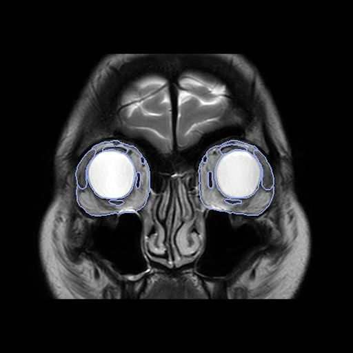
Coronal T2-weighted MRI of the orbits of a patient with thyroid eye disease at the first visit, with nine structures of both orbits outlined by hand (TOM500, Shanghai Ninth People's Hospital).
100 test casesorbitimage MR
Read before comparing (7)
- Cohort. 500 patients with clinically confirmed thyroid eye disease at their first visit, without another orbital disease, surgery or trauma. One scanner and one protocol, so the draw has no strata: Philips Ingenia 3.0 T, coronal T2-weighted turbo spin echo (T2-DRIVE), slices 3.5 mm thick and 3.85 mm apart, pixels of 0.3125 mm, 20 slices.
- Values. The files' own values after the provider's rescale, with no common scale: 16-bit integers on nine of the twenty images drawn first, 64-bit floats on the eleven others.
- Truth. Three junior orbital radiologists outlined the nine structures slice by slice in ITK-SNAP, blind to disease status; STAPLE fused their maps and an orbital radiology expert corrected the result. Each annotator scores a mean Dice of 0.95 against it, but that map is made from theirs and does not measure the truth's own error. The inferior oblique is not outlined.
- Labels. Both orbits share each id, hence the plural names. On the twenty images drawn first every structure lies where it should in both orbits, one piece an orbit but for at most 0.1 ml of stray voxels. The headers' axes are right where the labels can tell; left and right cannot be checked.
- Ignore. The protocol outlines each orbit from where its fat appears to where its muscles end, 8 to 11 of the 20 slices. The slices outside, orbit by orbit, are an ignore region never shown to methods: outlining the whole eyeball or optic nerve, or a muscle past the protocol's end, costs nothing.
- Size. The structures are small: on the 100 test cases, 23 to 49 ml of orbital fat, 1.7 to 4.8 ml of lacrimal gland, 0.7 to 1.7 ml of optic nerve and 0.5 to 1.7 ml of superior oblique. Across the slices a surface is known to 3.85 mm, against 0.31 mm pixels, so HD95 and the surface Dice at 1 mm move by whole slices.
- Seen. A network trained on the provider's suggested training split has not seen the test cases. The paper reports no network trained on TOM500, and TotalSegmentator makes none of the nine structures from MR.
No method has a result on every case of this dataset yet.
Case by case
Click a score to look at that result: the image, the truth in green and the method's map in red. Underlined scores have their image; for the others you see the case without the result.
About the data
The first test caseClick to look at every case.
- Test cases
- The 100 patients of the provider's validation split (split.json) are the test cases, taken whole. 30 development patients are drawn with seed 2026 from its 400 training patients, the last of the same seeded order. One scan a patient, so no patient is in both.
- Truth
- contours by experts the dataset's nine structures, both orbits under each id, outlined slice by slice by three junior orbital radiologists, fused by STAPLE, then reviewed and corrected by an orbital radiology expert; scored on the slices the protocol outlines in each orbit
- Source
- doi.org/10.6084/m9.figshare.27133389.v1
doi.org/10.1038/s41597-025-04427-9
github.com/guest0417/TOM500 - Licence
- CC0 1.0
30 cases for tuning, never tested
198, 1018, 281, 402, 1417, 439, 227, 1031, 385, 1149, 370, 386, 1386, 1026, 350, 283, 1071, 1139, 1576, 1106, 1120, 394, 1015, 1451, 1317, 1324, 1400, 422, 1344, 1600