 fidopt
Human inner ear CT, fluid spaces
Flat-panel volume CT at 0.0986 mm of one cadaver temporal bone, with the inner ear's fluid space labelled by hand: cochlea, vestibule, semicircular canals.
10 test caseshead and neckimage CT
Read before comparing (2)
- Cases. The fixed images of InnerEarCT's pairs: a test pair's image is a test case, a development pair's a development case. The moving images are left out: their headers carry the pair's rigid start, and the left ears among them are mirrored, so they are not the images as delivered.
- Donors. The dataset does not say which specimens come from one donor. The 39 right ears are 39 people; a left ear may be the other ear of one of them, so a mirrored pair may join the two ears of one person.
No method has a result on every case of this dataset yet.
Case by case
Click a score to look at that result: the image, the truth in green and the method's map in red. Underlined scores have their image; for the others you see the case without the result.
About the data
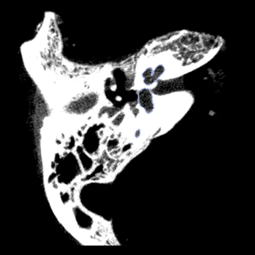
The first test caseClick to look at every case.
- Truth
- contours by experts the dataset's manual labels of the inner ear's fluid space: 1 cochlea, 2 vestibule, 3 semicircular canals
- Licence
- CC BY 4.0
10 cases for tuning, never tested
01_22/fixed, 29_12/fixed, 08_35/fixed, 05_19/fixed, 20_07/fixed, 09_37/fixed, 40_03/fixed, 34_02/fixed, 44_21/fixed, 06_18/fixed