 fidopt
CC-Tumor-Heterogeneity
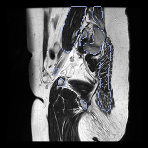
Sagittal T2 MR of the pelvis of one patient with advanced cervical cancer, before chemoradiotherapy (fixed) and 2 to 2.5 weeks into it (moving), from CC-Tumor-Heterogeneity (TCIA).
Read before comparing (9)
- Truth. The tumour shrinks between the scans: on the 20 cases its second contour holds 0.04 to 1.56 of the first's volume, median 0.57. No registration can make the two contours overlap, so the board ranks the distance between their centres, one point a case, which an even shrinkage keeps in place.
- Limits. Uneven regression moves the centre by an amount nobody measured, up to the first tumour's radius (13 to 39 mm on the test pairs). Part of what is left on a pair may be no registration error, and no method can be expected to reach zero.
- Contours. Each structure set holds two hand contours drawn in MIM, both named Ut-MRT2-Sag-<time>; the collection does not say which is which or who drew them. Read on the images, ROI 1 outlines the uterus, body and cervix, and ROI 2, 80 to 100% inside it, the tumour. Shown as dice_tumour and dice_uterus, never ranked.
- Labels. TotalSegmentator's organs, shown and not ranked: 19 to 25 structures on both images of a test pair, but no uterus, cervix or vagina, and its prostate taken out. Fragments at the field's edges (femurs, gluteus medius, the bottom of the aorta and vena cava) pull the mean Dice down. Where the uterus is distended its bladder runs into the experts' uterus contour, by 266 to 310 ml on test pair CCTH-B02.
- Alignment. Each session is in its scanner's frame, the tumour centres 17 to 146 mm apart as delivered. The moving image's, labels' and landmark's headers carry one elastix translation on mutual information inside each body, no label or contour used; both scans keep their voxels. It brings the bones together on every test pair looked at, the pelvis tilted by a few degrees on CCTH-A04.
- Baseline. With no registration the 10 test pairs score a centre error of 9.2 mm (3.8 to 18.8), a label Dice of 0.50 (0.27 to 0.64), an HD95 of 17.6 mm (10.4 to 26.2), a tumour Dice of 0.49 (0.14 to 0.81) and a uterus Dice of 0.65 (0.31 to 0.83).
- Images. The sagittal T2 series the contours were drawn on (spin echo, 1.5 T), both times on one scanner: GE Signa HDxt (CCTH-A01 to A10), Philips Ingenia (A11, A12), Siemens Espree (B01 to B11). The slab of 4 mm slices, 12 to 22.5 cm wide, cuts the hip bones and femurs differently each session.
- Times. The pair is the first two of three scans, before treatment and at 20 to 25 Gy, 16 to 38 days apart; every patient has both, the tumour still contoured at the second. The third time, the PET/CT, the other MR series and the provider's REG objects are not used.
- Outside numbers. None: the collection serves the study of tumour heterogeneity, not registration.
| Accuracy | Smoothness | Cost | ||||||||||
|---|---|---|---|---|---|---|---|---|---|---|---|---|
| # | Method | TRE mm | Change | Dice * | HD95 mm * | rTRE | SDlogJ | Folds % | Time s | RAM GB | GPU GB | Images |
| 1 | ConvexAdam (MIND)GPUapp | 8.94 ±4.90 | −0.21 | 0.614 ±0.056 | 15.61 ±3.52 | 0.0215 ±0.0117 | 0.090 | 0.29 | 9 | 2.2 | 1.3 | |
| 1 | DemonsCPUapp | 7.17 ±3.50 | −1.98 | 0.576 ±0.057 | 16.45 ±3.27 | 0.0172 ±0.0086 | 0.138 | 1.52 | 4 | 0.6 | ||
| No registration | 9.15 ±3.32 | 0.501 ±0.082 | 17.64 ±3.74 | 0.0218 ±0.0077 | 0.000 | 0.00 | ||||||
The order averages each method's ranks on TRE mm, SDlogJ, so the first one is not always the best in every column. ± is the 95% interval of the mean over the 10 pairs. Two neighbours are tied when a paired Wilcoxon test on the shared pairs does not find the upper one better on TRE mm (5%): the places a method could hold are then shown under its own. The best value of each column is in colour. Click a row to see its images, a column title to sort by it. Time and memory measured on Laptop.
* shown, not ranked: on this dataset it is checked against the output of a program.
Dice: how much the organ contours overlap, 1 is perfect. HD95: distance between the contours' surfaces. TRE: distance between matching landmarks. rTRE: the TRE over the length of the image's diagonal, to compare datasets of other sizes and units. SDlogJ: how irregular the deformation is, 0 is perfectly smooth. Folds: share of the image where the deformation folds over itself.
Pair by pair
Click a score to look at that result: the fixed image, and over it the moving image after registration, with the annotations. Underlined scores have their image; for the others you see the pair without the result. A green cell gained against no registration on that pair, a red one lost.
About the data
- Test pairs
- 10 test pairs drawn with seed 2026 by scanner, in proportion (4 GE, 1 Philips, 5 Siemens), among the 20 of the 23 patients outside the first three development pairs (CCTH-A01, A02 and B01); then 7 more development pairs, the last of the same seeded order. CCTH-A07, A12 and B08 are used for neither.
- Truth
- landmarks by experts contours by a program tumour by experts uterus by experts TotalSegmentator (task total_mr) run by us on each image, its prostate taken out: the bladder, the colon with the rectum, the small bowel, the sacrum, lumbar vertebrae, discs and spinal cord, the hip bones, the iliac vessels and the muscles of the pelvis and back the centre of the tumour on each image, the centroid of the experts' tumour contour (ROI 2) as filled on the image's voxels, one pair a case: 10 in the ten test pairs
- Source
- www.cancerimagingarchive.net/collection/cc-tumor-heterogeneity
doi.org/10.7937/ERZ5-QZ59
doi.org/10.1002/jmri.25874 - Licence
- CC BY 4.0
10 pairs for tuning, never tested
CCTH-A01, CCTH-A02, CCTH-B01, CCTH-B09, CCTH-A10, CCTH-B03, CCTH-A09, CCTH-B07, CCTH-A06, CCTH-B04