 fidopt
FDG-PET-CT-Lesions (autoPET), CT to PET
Contrast-enhanced CT (source) and FDG-PET in SUV (target) from one whole-body PET/CT session, neck down, of a patient with melanoma, lymphoma or lung cancer, or a negative control, on the PET's grid (FDG-PET-CT-Lesions, autoPET).
Read before comparing (11)
- Alignment. CT and PET come from one session on one Siemens Biograph mCT, on one table and in one frame of reference, the PET 3 to 31 minutes after the CT. Following the provider's conversion script, the CT is resampled onto the PET's grid by a cubic spline, -1024 outside the CT, and rounded to whole Hounsfield units. Nothing is registered.
- Target. Attenuation-corrected PET in body-weight SUV, the dose decayed to the start of the PET, as the provider's NIfTI release computes it since its version 2; the conversion script's own SUV, up to 20% higher, is not used. On PETCT_3049d4f1a4_2002-02-24 both images agree with that release, within 0.01 SUV and 0.02 HU.
- CT. The session's diagnostic CT, with a soft-tissue kernel, contrast-enhanced on all but 61 of the collection's 1,014; on the PET's grid it keeps no detail finer than those voxels. Where the PET's field goes past the CT's, the source reads -1024 and the body is left out of the mask.
- Motion. The CT takes 14 to 60 s, the PET 13 to 34 minutes. Breathing, bowel motion and the filling bladder make the two images disagree near the diaphragm, in the bowel and at the bladder by an amount nobody measured: there a perfect synthesis is still scored off, alike for every method.
- Uptake. FDG shows metabolism, which a CT shows only through anatomy. Uptake in the heart, bowel, muscles and brown fat varies between sessions, urine carries the tracer into the kidneys and bladder (up to 188 SUV), and 16 of the 30 test cases hold FDG-avid tumours: what the CT does not show counts against every method.
- Defacing. The public images are the collection's defaced version 2: from the base of the neck up, a box is blanked in each image, the two set separately, their lower edges 0 to 48 mm apart. The CT often shows the neck, shoulders or raised arms where the PET is blank.
- Mask. The body where both images hold it: the CT above -500 HU with the air it encloses in the body (lungs, airways), opened by one voxel to detach the table, its largest component, holes filled slice by slice, minus every voxel where the PET reads exactly 0. The target is scored in it, and methods are given it.
- Range. PSNR and SSIM take the SUV range 0 to 20, which holds the normal tissue and most tumours; urine in the bladder and kidneys and the hottest lesions lie past it.
- Baseline. On the 30 test cases a constant at the target's median inside the mask scores, as the median over the cases and its range, an MAE of 0.45 (0.36 to 0.63) SUV, a PSNR of 24.2 (14.8 to 29.4) dB and an SSIM of 0.75 (0.70 to 0.81).
- Overlap. AutoPETCT and AutoPET_Seg come from the same collection; their patients are left out, so that no patient is in two cases or two datasets.
- Outside numbers. None: autoPET is a segmentation challenge on these images.
No method has a result on every case of this dataset yet.
Case by case
Click a score to look at that result: the target image, and over it the method's synthetic image. Underlined scores have their image; for the others you see the case without the result.
About the data
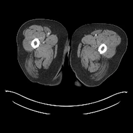
- Test cases
- 30 test studies drawn with seed 2026 by diagnosis: 6 melanoma, 5 lymphoma, 5 lung cancer and 14 negative. 30 development studies: the first study of each cancer group in alphabetical order, then the last of the same seeded order, those the test draw rests on kept, each diagnosis in proportion: 4 melanoma, 5 lymphoma, 6 lung cancer and 15 negative, none a patient of another dataset. The pool is each patient's first study, 778 of 900: left out are the 120 patients of AutoPETCT and AutoPET_Seg, PETCT_0223010e46, once an AutoPETCT development study, and PETCT_c5a143a604_2006-08-12, whose header gives no SUV. The strata's 880 studies already lack 20 of them; draw.held_out lists the other 102.
- Truth
- target image as released the FDG-PET of the same session, attenuation-corrected, in body-weight SUV, on its own grid, which the CT is brought onto through the scanner's frame of reference
- Source
- www.cancerimagingarchive.net/collection/fdg-pet-ct-lesions
doi.org/10.7937/gkr0-xv29
doi.org/10.1038/s41597-022-01718-3
github.com/lab-midas/TCIA_processing
doi.org/10.57754/FDAT.8f14a-pf846
github.com/medical-physics-usz/z-rad - Licence
- CC BY 4.0
30 cases for tuning, never tested
PETCT_01140d52d8_2005-08-13, PETCT_04ab5c61c9_2002-11-22, PETCT_01682f60c3_2002-03-11, PETCT_ca29501275_2004-11-04, PETCT_ded50b1e68_2006-02-09, PETCT_4bab91dccd_2006-01-05, PETCT_7cfaf65584_2006-10-28, PETCT_7c96c4626d_2003-06-30, PETCT_f15b868db6_2006-02-03, PETCT_d6a491e16d_2003-05-30, PETCT_5001354a69_2002-11-28, PETCT_72725f2bda_2007-07-06, PETCT_1fa22c576e_2004-10-17, PETCT_75e42ff05f_2002-09-21, PETCT_98cd91d44e_2003-09-06, PETCT_d64d6a87c8_2004-12-18, PETCT_e4e99dbb47_2005-07-17, PETCT_48cb8662f7_2003-03-07, PETCT_c58477f3b4_2002-12-07, PETCT_108c1763d4_2004-09-30, PETCT_a2a68bbe97_2003-08-21, PETCT_b258dfa7c2_2002-12-15, PETCT_16cdbc8689_2005-07-15, PETCT_d9da6129f8_2002-04-14, PETCT_d69eabbb20_2001-02-01, PETCT_44b08f570e_2001-02-10, PETCT_5d553bf6b4_2001-09-16, PETCT_60baa6979c_2003-08-16, PETCT_44d6ba6772_2003-07-13, PETCT_bd52fdf529_2000-12-03