 fidopt
PSMA-PET-CT-Lesions (autoPET III and IV), CT to PET
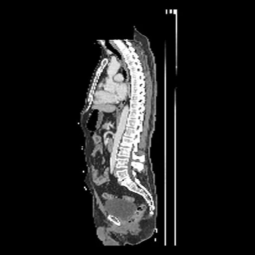
One whole-body PSMA-PET/CT session of a man with suspected or diagnosed prostate cancer: the diagnostic CT brought onto the PET's grid (source) and the PET in SUV (target) (PSMA-PET-CT-Lesions, autoPET III and IV).
Read before comparing (11)
- Cohort. 597 studies of 378 men before and/or after therapy, at LMU University Hospital Munich, with 18F-PSMA or 68Ga-PSMA.
- Alignment. The CT and the PET of one session share one table and one frame of reference; the PET starts 0.7 to 2.0 minutes after the CT's first slice. The CT is resampled onto the PET's grid by a cubic spline, as the provider's recipe does (lab-midas/TCIA_processing), rounded to whole Hounsfield units. Nothing is registered.
- Target. The attenuation-corrected PET in body-weight SUV, the dose decayed to the start of the PET, as the provider's NIfTI release computes it. On four studies compared with that release the SUV agrees within 0.003 SUV and the CT within 0.5 HU, except at the GE reconstructed circle, the edge of the CT's field and the defaced boxes.
- Slices. The CT of development case PSMA_738bc5d9946240f3_1997-09-26 is unevenly sliced; spread evenly, its slices would lie up to 18 cm off. prepare.py fills any such CT in linearly on a 2.5 mm grid before the spline.
- Motion. The CT takes seconds, the PET 12 to 31 minutes. Breathing, bowel motion and the filling bladder make the two images disagree near the diaphragm, in the bowel and at the bladder by an amount nobody measured: a perfect synthesis is still scored off there, the same for every method.
- Uptake. PSMA uptake follows a membrane protein that the CT shows only through anatomy: kidneys past 20 SUV on most cases, tracer in the urine, uptake in liver, spleen and bowel that varies with the case and the tracer, and lesions. What the CT does not show counts as error for every method.
- Mask. The body where both images hold it: the CT above -500 HU, lungs and airways included, the table detached and holes filled, less every voxel where the PET reads exactly 0. TCIA's defacing blanks a box over the skull base and neck separately on each image, so the CT may show the neck or arms where the PET is blank. The target is scored in the mask, which the method is given.
- Range. truth.dynamic_range is 20, as in AutoPET_CTPET_Syn, so both tracers' PSNR and SSIM read on one SUV scale. Kidneys, urine and the hottest lesions reach 377 SUV in the test masks, a case's hottest voxel 71 SUV at the median; nothing is clipped, so those voxels count with their whole error.
- Baseline. On the 24 test cases a constant at the target's median inside the mask scores, as the median over the cases and its range, an MAE of 0.80 (0.53 to 1.12) SUV, a PSNR of 16.7 (11.8 to 21.9) dB and an SSIM of 0.73 (0.61 to 0.77).
- Held out. PSMA_e3c4639102fe5c51_2003-08-19 puts the PET 7.2 minutes after the injection, against 74 on average, while its image shows late uptake: the injection time is most likely wrong and no SUV can be computed. The 46 studies built pass that check.
- Overlap. No patient here is in AutoPSMA, PSMAFollowUp or PSMALesionsPET, made from the same collection; a later dataset of it should leave these patients out too. autoPET III and IV are segmentation challenges: no outside number compares.
No method has a result on every case of this dataset yet.
Case by case
Click a score to look at that result: the target image, and over it the method's synthetic image. Underlined scores have their image; for the others you see the case without the result.
About the data
- Test cases
- 24 test studies drawn by scanner with seed 2026 from a pool of 248 (9 Biograph mCT Flow 20, 9 Discovery 690, 6 Biograph 64-4R TruePoint), then 22 development studies: each scanner's first study in alphabetical order, then the last of the same seeded order, those the test draw rests on kept, each scanner in proportion (8, 11 and 3), none a patient of another dataset. The pool holds each patient's first study, less the 129 patients of AutoPSMA, PSMAFollowUp and PSMALesionsPET and one study with no usable SUV: the strata's 337 studies already lack 40 of those patients and that study, and draw.held_out lists the other 89.
- Truth
- target image as released the PSMA-PET of the same session, attenuation-corrected, in body-weight SUV, on its own grid, which the CT is brought onto through the scanner's frame of reference
- Source
- www.cancerimagingarchive.net/collection/psma-pet-ct-lesions
doi.org/10.7937/r7ep-3x37
www.cancerimagingarchive.net/wp-content/uploads/PSMA-PET-CT-Lesions-CLINICAL_v01_20251118.tsv
services.cancerimagingarchive.net/nbia-api/services/v1/getSeries?Collection=PSMA-PET-CT-Lesions
github.com/lab-midas/TCIA_processing
fdat.uni-tuebingen.de/records/rdkqd-wdh87
github.com/medical-physics-usz/z-rad - Licence
- CC BY 4.0
22 cases for tuning, never tested
PSMA_04f0f08528c9c231_2000-11-23, PSMA_02826eb561d6e0c7_1999-09-28, PSMA_0878fdec425f09c3_2002-04-16, PSMA_5c0552e946050837_2002-10-15, PSMA_7f274800860fcbfa_1998-05-22, PSMA_738bc5d9946240f3_1997-09-26, PSMA_9b7dfd431faf4067_2002-06-19, PSMA_dc8bb6a76a0a72cd_1999-09-24, PSMA_e8c1c3c25d7a735b_2000-06-22, PSMA_367adbcbbe2108d5_2002-08-20, PSMA_7fa68d5b0861c0b4_1997-09-22, PSMA_790ee2c577d4ceed_2003-10-01, PSMA_ada3f9f15facc495_2001-12-11, PSMA_43324b58ec80a0a5_2001-06-15, PSMA_ded97a4f2275ba94_2001-03-07, PSMA_e1eef1b5e130ded8_1999-06-02, PSMA_efb421c79f1368f9_1999-07-28, PSMA_0f434739488a6988_2000-09-26, PSMA_dee1112c5d1380f1_2001-08-29, PSMA_32e70fdbf4468564_2003-04-30, PSMA_46fcda50b67c2523_2003-10-15, PSMA_8e7619b9167225fb_1999-05-11