 fidopt
CT follow-up of liver metastases
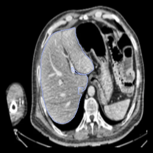
Portal-venous abdominal CTs of one patient with liver metastases, before and after systemic chemotherapy, 32 to 287 days apart (Al-Zahra Hospital, Isfahan).
Read before comparing (6)
- Values. Not Hounsfield units: the provider clipped each CT to level 40, width 450 HU, and scaled it to [0, 1], so a value v stands for about 450 v - 185 HU; the corpus keeps them. Fixed HU windows or thresholds and networks trained on HU see an almost flat image unless the method rescales. Air, lung, gas, bone and dense contrast are flat: nothing to register there.
- Scale. The headers lost the voxel size: every image says 1 mm, origin at zero, where the scans had 0.5 to 1.0 mm pixels and 1 to 1.2 mm slices. HD95 and fields are in the fixed image's voxels, and a pair's exams differ in scale: ANTs' affine stage finds factors of 0.49 to 1.50 along the body axis on the 12 built pairs where it works. Ranked apart for that, with LiverPhasesCT.
- Alignment. Each pair gets one elastix translation from the intensities, no annotation used, written in the moving image's header; nothing is resampled. The scale difference, up to a factor of two along the body axis, is left to the methods, so the board measures that as much as the liver's change.
- Baseline. With no registration the ten test pairs start at a mean liver Dice of 0.73 (0.24 to 0.86). The provider removed the slices without liver, so most images hold little more; Patient-49's second exam also holds the chest, and that pair starts lowest.
- Truth. Only the liver is scored, the metastases inside label 1 as the provider's liver mask holds them. They shrink, grow, appear or vanish under chemotherapy, so where they changed even a perfect field of the parenchyma does not reach a Dice of 1; the liver itself may change too.
- Masks. Drawn by a network, then corrected by hand and verified by radiologists. Some files mark a few voxels 2, a few metastasis voxels lie just outside the liver mask, and in Patient-02's first exam the two files are swapped: label 1 is every voxel either mask marks.
| Accuracy | Smoothness | Cost | ||||||||
|---|---|---|---|---|---|---|---|---|---|---|
| # | Method | Dice | Change | HD95 mm | SDlogJ | Folds % | Time s | RAM GB | GPU GB | Images |
| 11–2 | ConvexAdam (MIND)GPUapp | 0.863 ±0.118 | +0.129 | 18.92 ±15.24 | 0.113 | 0.87 | 12 | 2.7 | 3.5 | |
| 21–2 | DemonsCPUapp | 0.846 ±0.117 | +0.112 | 21.79 ±15.07 | 0.066 | 0.02 | 10 | 1.8 | ||
| No registration | 0.734 ±0.134 | 28.00 ±14.92 | 0.000 | 0.00 | ||||||
The order averages each method's ranks on Dice, HD95 mm, SDlogJ, so the first one is not always the best in every column. ± is the 95% interval of the mean over the 10 pairs. Two neighbours are tied when a paired Wilcoxon test on the shared pairs does not find the upper one better on Dice (5%): the places a method could hold are then shown under its own. The best value of each column is in colour. Click a row to see its images, a column title to sort by it. Time and memory measured on Laptop.
Dice: how much the organ contours overlap, 1 is perfect. HD95: distance between the contours' surfaces. SDlogJ: how irregular the deformation is, 0 is perfectly smooth. Folds: share of the image where the deformation folds over itself.
Pair by pair
Click a score to look at that result: the fixed image, and over it the moving image after registration, with the annotations. Underlined scores have their image; for the others you see the pair without the result. A green cell gained against no registration on that pair, a red one lost.
About the data
- Test pairs
- 10 test pairs drawn with seed 2026 among the 44 patients outside development, out of 54, and 10 development pairs. The earlier exam is the fixed image; a patient with three exams gives the pair of its first two, so no exam is scored twice.
- Truth
- contours by experts the liver (label 1), metastases included: the dataset's liver and metastasis masks together, drawn by a deep-learning network, corrected in ITK-SNAP by trained annotators and verified by radiologists
- Licence
- CC0 1.0
10 pairs for tuning, never tested
Patient-01, Patient-02, Patient-03, Patient-20, Patient-26, Patient-07, Patient-05, Patient-29, Patient-13, Patient-22