 fidopt
Baby Open Brains (BOBs)
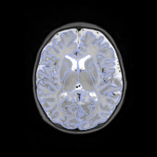
Brain T2w MR of one infant at two visits 2 to 6 months apart, between 1 and 9 months of age (Baby Open Brains, from the Baby Connectome Project).
Read before comparing (9)
- Truth. Each visit's map was started by a program (joint label fusion, then BIBSNet), corrected by hand in ITK-SNAP by trained markers and checked by at least one expert. The two maps of a pair are two readings, and no agreement between markers is published. BIBSNet was trained on these maps: a method built on its labels or features is scored on its training data.
- Labels. The structures the protocol has the markers edit, and the ventral diencephalon: 31 labels on both visits of every pair (28, the left ventral diencephalon, is missing from the dataset's dseg.tsv). prepare.py sets the others to 0: vessels, choroid plexus, CSF, white matter hypointensities, optic chiasm, vermis and the cerebral and cerebellar exteriors, which come and go between visits.
- Contrast. The T2w is taken because its contrast holds better between visits: it keeps the sign of white matter against cortex within 7 of the 10 test pairs, the T1w within 4. Inside the brain the two aligned T2w still correlate only between -0.19 and 0.41: a metric that expects the same intensities, SSD or NCC, meets a changed contrast.
- Growth. The labelled brain grows by 17 to 63% within a test pair, unevenly: by a median 43% for the cortex, 64% for the cerebellum, 23% for the white matter and 0% for the thalamus; the lateral ventricles shrink by a median 8%. A correct field changes volume, so its Jacobian columns do not read as on an adult pair.
- Ceiling. A perfect registration leaves an overlap below 1: two readings at two stages of myelination, which differ most at the cortical folding, the unmyelinated white matter and the subcortical boundaries. New tissue has no point to match: the labels score whether each structure lands on its counterpart.
- Alignment. Both visits come on one 1 mm INFANTMNIacpc grid but are not aligned (Dice 0.06 to 0.71 as delivered). The moving image's and labels' headers carry one elastix rigid transform on mutual information inside each head, no label used; both visits keep their voxels. A translation alone left sub-584381 tilted by 22 degrees.
- Baseline. With no registration the ten test pairs start at a mean Dice of 0.596 (0.387 to 0.745) and a mean HD95 of 4.39 mm (2.28 to 7.42).
- Images. One site (University of Minnesota) and one scanner (Siemens 3 T Prisma). Defaced, not skull-stripped. No mask is given: one taken from the labels would hand over the truth.
- Version. The bucket holds version 1.0; the providers keep correcting the segmentations, and `fidopt verify` tells a later release from this one.
| Accuracy | Smoothness | Cost | ||||||||
|---|---|---|---|---|---|---|---|---|---|---|
| # | Method | Dice | Change | HD95 mm | SDlogJ | Folds % | Time s | RAM GB | GPU GB | Images |
| 1 | ConvexAdam (MIND)GPUapp | 0.762 ±0.031 | +0.166 | 2.68 ±0.56 | 0.071 | 0.20 | 9 | 2.4 | 2.1 | |
| 2 | DemonsCPUapp | 0.719 ±0.047 | +0.123 | 2.88 ±0.60 | 0.051 | 0.00 | 6 | 1.1 | ||
| No registration | 0.596 ±0.093 | 4.39 ±1.29 | 0.000 | 0.00 | ||||||
The order averages each method's ranks on Dice, HD95 mm, SDlogJ, so the first one is not always the best in every column. ± is the 95% interval of the mean over the 10 pairs. Two neighbours are tied when a paired Wilcoxon test on the shared pairs does not find the upper one better on Dice (5%): the places a method could hold are then shown under its own. The best value of each column is in colour. Click a row to see its images, a column title to sort by it. Time and memory measured on Laptop.
Dice: how much the organ contours overlap, 1 is perfect. HD95: distance between the contours' surfaces. SDlogJ: how irregular the deformation is, 0 is perfectly smooth. Folds: share of the image where the deformation folds over itself.
Pair by pair
Click a score to look at that result: the fixed image, and over it the moving image after registration, with the annotations. Underlined scores have their image; for the others you see the pair without the result. A green cell gained against no registration on that pair, a red one lost.
About the data
- Test pairs
- 10 test pairs drawn with seed 2026 among the 14 outside the first three development pairs, and 7 development pairs: every infant with two visits or more, 17 of the 51, one pair each. The three infants with three visits give their first and last.
- Truth
- contours by experts the dataset's segmentation of each visit, 31 structures: cerebral white matter and cortex, ventricles, cerebellum, deep grey nuclei, hippocampus, amygdala, ventral diencephalon and brainstem
- Source
- registry.opendata.aws/bobsrepository
doi.org/10.1038/s41597-025-05404-y
doi.org/10.17605/OSF.IO/WDR78
openneuro.org/datasets/ds005450
bobsrepository.readthedocs.io - Licence
- CC BY 4.0
7 pairs for tuning, never tested
sub-116845_ses-3mo_ses-6mo, sub-198202_ses-5mo_ses-8mo, sub-198549_ses-1mo_ses-7mo, sub-470437_ses-1mo_ses-7mo, sub-272097_ses-5mo_ses-8mo, sub-710922_ses-4mo_ses-7mo, sub-381606_ses-2mo_ses-8mo