 fidopt
CPTAC-UCEC-Tumor-Annotations
A post-contrast T1-weighted pelvic MR of a woman with endometrial carcinoma before treatment, the uterus holding the tumour outlined as a target lesion by radiologists and reviewed by board-certified radiologists (CPTAC-UCEC-Tumor-Annotations on CPTAC-UCEC, TCIA).
10 test casespelvisimage MR
Read before comparing (9)
- Annotations. Version 2 of 2025-10-20 of TCIA's annotations, public since 2023. Radiologists holding MBBS degrees or higher drew each lesion by hand in the axial plane, on all axial T1-weighted post-contrast sequences, and US board-certified radiologists reviewed it, as RECIST 1.1 target lesions: five a scan and two an organ at most.
- Cases. A case is a patient's MR series with a contour named 'UTERUS', before treatment: the earliest such examination, then the series with the most images, then the highest series number; of a dynamic acquisition, the last phase. 34 patients have one: the pool. 30 of the 34 series are one Siemens Avanto_fit protocol, those naming no maker included, so the strata are makers as the series give them, not sites.
- Truth. On 18 of the 20 cases built the outline follows the outer wall of the uterus, with the myometrium and the tumour inside; on C3L-01248 (test) and C3N-01764 (development) it encloses a region inside the wall. The label is named uterus: a method that segments the tumour alone is charged the myometrium, and on those two cases a method that segments the uterus is charged the wall.
- Ranked apart. The truth is people's and reviewed but not one structure throughout, so the set misses the bar of a main leaderboard; a tumour set would need the contours redrawn. TotalSegmentator's total_mr makes no uterus, so no network checked the label.
- Not labelled. The caps leave the pelvic and iliac nodes, the ovaries and the pelvic masses beyond them undrawn. Only the uterus' contours are written, one on 32 of the 34 series and two on 2; the other target lesions, on 6 series, are not. The 34 patients annotated on CT alone are not built: CT shows the tumour poorly.
- Images. 18 of the 20 series built follow the Siemens protocol, 0.80 to 1.03 mm in plane, slices 2 mm apart. C3L-01248 (test, GE) has 20 slices 10 mm apart, four of them labelled; C3L-00963 (development, GE) is sagittal, the contours drawn in its plane. The values are the scanner's, on scales that differ (maxima of 415 to 3512).
- Volumes. The ten test labels hold 32 to 583 ml (median 82; 16 to 252 ml in development), in one piece but C3N-00858 (two). The filled volumes agree with the report's on 17 cases (0.97 to 1.16 times); the report gives 93.5 ml for C3N-02595's 252, 7.8 ml for C3N-01764's 15.9 and 0.24 ml for C3L-00963's 41.5.
- Room. Moved by one voxel in plane, the test masks keep a Dice of 0.93 to 0.98 (mean 0.97); by one slice, 0.58 (C3L-01248, 10 mm slices) to 0.96 (0.90).
- Overlap. SAROSBodyCT holds CTs of 7 other CPTAC-UCEC patients and leaves these 20 out. None of the 40 images TotalSegMR and LiverSegmentsMR, the corpus's sets of Imaging Data Commons scans, then held matches an MR series of CPTAC-UCEC by slice count, rows, columns and pixel size; a series holding several volumes was not tried. UterineMyomaMR, the other uterus set, holds sagittal T2 of other women.
No method has a result on every case of this dataset yet.
Case by case
Click a score to look at that result: the image, the truth in green and the method's map in red. Underlined scores have their image; for the others you see the case without the result.
About the data
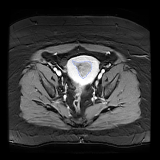
The first test caseClick to look at every case.
- Test cases
- 10 test patients drawn with seed 2026 by the maker of the annotated MR among the 24 outside development (5 Siemens, 4 whose series names no maker, 1 GE), and 10 development patients, the last of the same seeded order; one series each.
- Truth
- contours by experts the target lesion named 'UTERUS', drawn by hand in the axial plane by radiologists (MBBS or higher) and reviewed by US board-certified radiologists: on 18 of the 20 cases built the outline follows the outer wall of the uterus that holds the tumour, on two (C3L-01248, a test case, and C3N-01764) a region inside it
- Source
- www.cancerimagingarchive.net/analysis-result/cptac-ucec-tumor-annotations
doi.org/10.7937/89M3-KQ43
www.cancerimagingarchive.net/wp-content/uploads/Metadata_Report_CPTAC_UCEC_2025-10-20.csv
www.cancerimagingarchive.net/collection/cptac-ucec
doi.org/10.7937/K9/TCIA.2018.3R3JUISW
services.cancerimagingarchive.net/nbia-api/services/v1/getSeries?Collection=CPTAC-UCEC - Licence
- CC BY 4.0
10 cases for tuning, never tested
C3N-01007, C3N-01342, C3N-01172, C3N-03417, C3N-01003, C3N-02979, C3N-02595, C3N-01877, C3N-01764, C3L-00963