 fidopt
RESECT
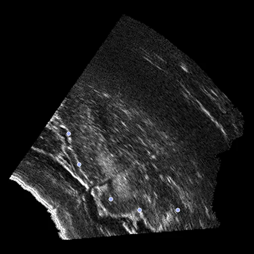
Intra-operative ultrasound before resection (fixed) and pre-operative FLAIR MR (moving) of one patient with a low-grade glioma (RESECT, the data of the CuRIOUS challenges).
Read before comparing (2)
- Alignment. Both images come in the frame of the neuronavigation system, the alignment a surgeon starts from: 1 to 20 mm off at the landmarks.
- Field. The ultrasound covers part of the brain around the tumour, and the field is asked on its grid. The mask is where the sweep recorded something.
No method has a result on every pair of this dataset yet.
| Accuracy | Smoothness | Cost | ||||||||
|---|---|---|---|---|---|---|---|---|---|---|
| # | Method | TRE mm | Change | rTRE | SDlogJ | Folds % | Time s | RAM GB | GPU GB | Images |
| No registration | 4.83 ±2.27 | 0.0371 ±0.0175 | 0.000 | 0.00 | ||||||
TRE: distance between matching landmarks. rTRE: the TRE over the length of the image's diagonal, to compare datasets of other sizes and units. SDlogJ: how irregular the deformation is, 0 is perfectly smooth. Folds: share of the image where the deformation folds over itself.
Pair by pair
Click a score to look at that result: the fixed image, and over it the moving image after registration, with the annotations. Underlined scores have their image; for the others you see the pair without the result. A green cell gained against no registration on that pair, a red one lost.
About the data
- Test pairs
- 10 test pairs drawn with seed 2026 among the 12 training patients outside development, and 10 development pairs. The challenge's test landmarks are not public, so all pairs come from its 22 training patients.
- Truth
- landmarks by experts the dataset's expert landmark pairs, 15 or 16 a patient
- Licence
- CC BY 4.0
10 pairs for tuning, never tested
case01, case02, case03, case18, case21, case26, case25, case14, case12, case19