 fidopt
Prostate-MRI-US-Biopsy
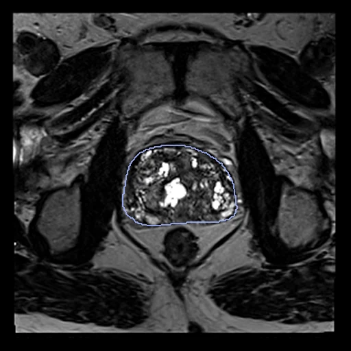
T2 MR (fixed) and 3D transrectal ultrasound (moving) of one patient's prostate, the MR before an MR-ultrasound fusion biopsy and the ultrasound at its start (Prostate-MRI-US-Biopsy, UCLA).
Read before comparing (7)
- Truth. Semi-automatic gland contours: the user traced the prostate on one or more planes, the software segmented the rest in 3D, and the user could move its vertices; TCIA finds them generally good, their accuracy depending on the user. A pair's two contours were drawn apart with two programs, so no registration reaches a Dice of 1: the ultrasound gland is 14% smaller to 46% larger than the MR's on the test pairs (16% larger at the median).
- Left out. The ultrasound's lesion regions and the biopsy cores' MR coordinates rest on the vendor's nonrigid fusion, 3 to 4 mm on average according to TCIA: scored against them, a method would be scored against that registration.
- Geometry. The ultrasound's DICOM holds no geometry: prepare.py gives it the manufacturer's convention as SlicerHeart reads it, isotropic voxels of 0.25 to 0.44 mm centred on 0, turned by the axis permutation TCIA applied to the ultrasound surfaces, which then lie on the gland on the 13 pairs checked by eye.
- Alignment. The two images come on unrelated grids. The start puts their geometric centres together (image-centres), nothing optimised and no contour used, in the moving image's and gland's headers. An elastix translation on intensities left a Dice of 0.17, 0.00 on ten pairs, and is not used. The probe's angle and the deformation are left to the methods.
- Baseline. With no registration the 30 test pairs start at a gland Dice of 0.62 (0.40 to 0.80) and an HD95 of 12.8 mm.
- Patients. 257 of the 842 patients have more than one pair; only the first biopsy is used. The MR came 2 to 193 days before the ultrasound on the test pairs. Test pair 0289's MR is a GE scan with an endorectal coil, one of the few TCIA says came from other institutions; the rest are UCLA's 3 T Siemens scans.
- Contours. The MR contours filled here match the Imaging Data Commons' DICOM SEG of the same surfaces at a Dice of 0.996 to 0.997 on the 13 pairs checked. The ultrasound surfaces are only in TCIA's STL package, behind a public link that needs no account.
| Accuracy | Smoothness | Cost | ||||||||
|---|---|---|---|---|---|---|---|---|---|---|
| # | Method | Dice | Change | HD95 mm | SDlogJ | Folds % | Time s | RAM GB | GPU GB | Images |
| 1 | ConvexAdam (MIND)GPUapp | 0.621 ±0.050 | +0.002 | 15.06 ±1.49 | 0.253 | 5.93 | 7 | 2.3 | 1.4 | |
| 2 | deedsCPUapp | 0.514 ±0.066 | −0.105 | 26.40 ±3.59 | 0.155 | 4.64 | 15 | 0.8 | ||
| 3 | NiftyRegCPUapp | 0.299 ±0.065 | −0.320 | 36.80 ±3.53 | 1.458 | 7.75 | 19 | 1.3 | ||
| No registration | 0.619 ±0.042 | 12.85 ±1.30 | 0.000 | 0.00 | ||||||
The order averages each method's ranks on Dice, HD95 mm, SDlogJ, so the first one is not always the best in every column. ± is the 95% interval of the mean over the 30 pairs. Two neighbours are tied when a paired Wilcoxon test on the shared pairs does not find the upper one better on Dice (5%): the places a method could hold are then shown under its own. The best value of each column is in colour. Click a row to see its images, a column title to sort by it. Time and memory measured on Laptop.
Dice: how much the organ contours overlap, 1 is perfect. HD95: distance between the contours' surfaces. SDlogJ: how irregular the deformation is, 0 is perfectly smooth. Folds: share of the image where the deformation folds over itself.
Pair by pair
Click a score to look at that result: the fixed image, and over it the moving image after registration, with the annotations. Underlined scores have their image; for the others you see the pair without the result. A green cell gained against no registration on that pair, a red one lost.
About the data
- Test pairs
- 30 test patients drawn with seed 2026, 10% of the 842 with a contoured MR and ultrasound pair, at most 30, then 30 development patients from the 812 left. A case is the pair of each patient's first biopsy.
- Truth
- contours by a program, verified by people the prostate gland on each image, contoured semi-automatically with Eigen's tools (ProFuse on the MR, the Artemis system on the ultrasound), filled by us on each image's grid
- Source
- www.cancerimagingarchive.net/collection/prostate-mri-us-biopsy
doi.org/10.7937/TCIA.2020.A61IOC1A
doi.org/10.1016/j.juro.2012.08.095
github.com/SlicerHeart/SlicerHeart/blob/master/Docs/ImageImportExport.md#eigen-artemis
zenodo.org/records/10069911 - Licence
- CC BY 4.0
30 pairs for tuning, never tested
Prostate-MRI-US-Biopsy-0001, Prostate-MRI-US-Biopsy-0002, Prostate-MRI-US-Biopsy-0003, Prostate-MRI-US-Biopsy-0321, Prostate-MRI-US-Biopsy-0459, Prostate-MRI-US-Biopsy-1138, Prostate-MRI-US-Biopsy-1000, Prostate-MRI-US-Biopsy-0653, Prostate-MRI-US-Biopsy-0517, Prostate-MRI-US-Biopsy-0914, Prostate-MRI-US-Biopsy-0713, Prostate-MRI-US-Biopsy-0908, Prostate-MRI-US-Biopsy-1037, Prostate-MRI-US-Biopsy-0075, Prostate-MRI-US-Biopsy-0876, Prostate-MRI-US-Biopsy-0309, Prostate-MRI-US-Biopsy-0668, Prostate-MRI-US-Biopsy-0650, Prostate-MRI-US-Biopsy-0152, Prostate-MRI-US-Biopsy-1004, Prostate-MRI-US-Biopsy-1038, Prostate-MRI-US-Biopsy-0421, Prostate-MRI-US-Biopsy-0718, Prostate-MRI-US-Biopsy-0455, Prostate-MRI-US-Biopsy-0802, Prostate-MRI-US-Biopsy-1067, Prostate-MRI-US-Biopsy-1111, Prostate-MRI-US-Biopsy-1142, Prostate-MRI-US-Biopsy-0328, Prostate-MRI-US-Biopsy-0425