 fidopt
4D-Lung, CBCT to CT
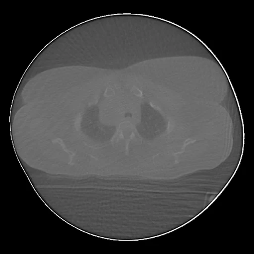
The 50% breathing phase of a lung cancer patient's 4D cone-beam CT at a radiotherapy session (source), registered by the corpus onto the same phase of their 4D fan-beam CT (target), from TCIA 4D-Lung.
Read before comparing (8)
- Cone beam. Each phase is made from about 250 projections: it carries view-aliasing streaks and little soft-tissue contrast, a quality the paper says may not be the clinic's. It covers 15 cm along the body, in a disc of 45 cm that cuts the shoulders, arms or flanks of larger patients (106 in test). Patient 113's spinal hardware streaks the whole image.
- Pairs. A patient's first 4D CT goes with the nearest cone-beam session, its last with the nearest other one; for 103, 104 and 106 one 4D CT serves twice. The images are 1 to 61 days apart: the tumour shrinks and the lung opens or collapses, which no registration undoes and which counts as error for every method. The ten test cases are not independent.
- Registration. The CT is never resampled: it is cut to the box the cone-beam field reaches, voxels untouched, and the cone-beam CT is resampled into it, linearly, through one registration: a rigid fit on mutual information, then IMPACT-Reg 1.8.7's preset Elastix_IMPACT_Jacobian with its defaults, ending in a cubic B-spline with a 10 mm grid. prepare.py rebuilds the same files from transforms/<case>.npz without registering.
- Check. TotalSegmentator ran on both images, the lungs, vertebrae and ribs each one structure: over the twenty cases the median mean Dice rises from 0.57 after the rigid start to 0.65 after registration (0.52 to 0.62 on the test cases), the lungs by threshold, owing the network nothing, from 0.72 to 0.85 (0.81 to 0.92 on the test cases). Every case gains on both, and none was dropped. These labels are not part of the dataset.
- Weak check. The network finds little on patient 113's streaked cone-beam CTs, where the lungs by threshold still gain and the overlays show the lungs, chest wall and diaphragm in place. Elsewhere a Dice of 0.58 to 0.80 is as much the network's reading of a streaked image as the registration's error.
- Mask. The CT's body, couch out and lungs in, inside the cone-beam field as the transform carries it: the disc of 250 pixels (220 mm) about each slice's centre, without the bright ring of its last 7 pixels. It holds 7.8 to 13.3 litres on the test cases.
- Baseline. The cone-beam values are not calibrated Hounsfield units: inside the mask they read 48 to 113 below the CT on the test cases, air about -1150. There a constant at the CT's median is 222 HU from the CT on average (138 to 296), the registered cone-beam CT 143 HU (112 to 186): a method must at least remove the offset and the streaks.
- Overlap. L2R_ThoraxCBCT's ten patients were matched by rigid registration of their cone-beam CTs: a correlation of 0.80 to 0.996 with one patient, under 0.72 with every other. Lung4DCT_Seg shares these patients and this partition, and gives every method the 0% or 50% phase of the 4D CT a target here comes from. The collection has no other free patient; the overlap is known and the dataset ranked apart.
No method has a result on every case of this dataset yet.
Under a licence that is not open
Methods whose code or weights are released for non-commercial use, or under share-alike or custom terms: shown for reference, ranked nowhere.
| Accuracy | Cost | ||||||||
|---|---|---|---|---|---|---|---|---|---|
| # | Method | MAE | PSNR dB | SSIM | Time s | RAM GB | GPU GB | Machine | Images |
| KonfAI synthesis app · TotalSynth CBCT to CT, KonfAI appGPUapptrained on CC-BY-NC-4.0 dataolder run | 88.2 ±16.3 | 29.91 ±1.59 | 0.636 ±0.099 | 30 | 3.7 | 4.4 | Workstation | ||
| nnsyn · CBCT to CT, thoraxGPUapptrained on CC-BY-NC-4.0 dataolder run | 66.8 ±8.7 | 31.80 ±1.43 | 0.736 ±0.071 | 3.9 | 2.6 | Laptop | |||
MAE: mean absolute error of a synthetic image against its target, inside the body, in the image's units. PSNR: the same by signal-to-noise ratio, higher is better. SSIM: the same by structural similarity, 1 is perfect.
Case by case
| MAE | 1 | 2 | 3 | 4 | 5 | 6 | 7 | 8 | 9 | 10 |
|---|
Click a score to look at that result: the target image, and over it the method's synthetic image. Underlined scores have their image; for the others you see the case without the result.
About the data
- Test cases
- 10 test cases from five patients and 10 development cases from five others, two sessions a patient: the first and the last five, in the order of sha256("2026:4D-Lung:<patient>"), of the 10 patients L2R_ThoraxCBCT does not hold. The partition is Lung4DCT_Seg's.
- Truth
- target image by a program the 50% phase of a 4D fan-beam CT of the patient in Hounsfield units, cut to the box of its grid the CBCT's field of view reaches; the CBCT's 50% phase registered onto it by the corpus with IMPACT-Reg 1.8.7 (Elastix_IMPACT_Jacobian, from a rigid start on mutual information)
- Source
- www.cancerimagingarchive.net/collection/4d-lung
doi.org/10.7937/K9/TCIA.2016.ELN8YGLE
doi.org/10.1002/mp.12059 - Licence
- CC BY 3.0
10 cases for tuning, never tested
107_2, 103_2, 115_2, 118_1, 118_2, 107_1, 104_2, 103_1, 104_1, 115_1