 fidopt
RIDER Lung CT
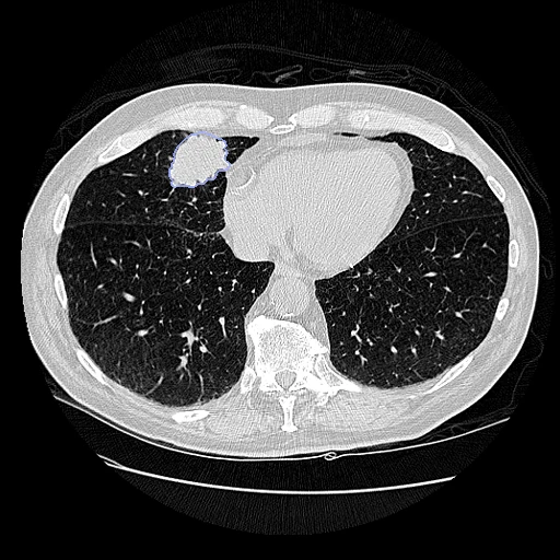
Chest CT of one patient with non-small cell lung cancer, scanned twice within 15 minutes on the same scanner, with the primary tumour drawn by hand on both (RIDER Lung CT, test and retest).
Read before comparing (5)
- Scans. Same scanner and protocol, each in one breath hold, 1.25 mm slices, lung kernel, no contrast; between them the patient left the table, walked around and lay down again.
- Alignment. The scans keep their native grids. The moving image's and labels' headers carry a rigid start from CT intensities and body masks alone, never the tumour, turning by 1.03 to 5.68 degrees over the 20 patients. prepare.py first sets the padding around the reconstructed circle, -3024 HU, to air.
- Baseline. On the ten test pairs the rigid start leaves a median tumour Dice of 0.778 and an HD95 of 5.58 mm, against 0.609 and 7.24 mm for a translation alone. The Dice improves on all ten; the HD95 rises by 0.78 and 1.00 mm on two.
- Truth. One radiation oncologist drew the primary tumour on each scan, blind to the other delineations; the SEG's automatic contour is not used. The two contours of a tumour differ in volume by 6 to 23%, and on RIDER-1760553574 their centres stay 9 mm apart while the anatomy around them matches: a perfect registration still leaves the contouring.
- Scored. Only the tumour counts, 0.8 to 92 ml, and on the small ones a millimetre moves the Dice a lot. The board says nothing of the rest of the chest, so this near-identity task stands apart from the main registration board.
| Accuracy | Smoothness | Cost | ||||||||
|---|---|---|---|---|---|---|---|---|---|---|
| # | Method | Dice | Change | HD95 mm | SDlogJ | Folds % | Time s | RAM GB | GPU GB | Images |
| 11–2 | greedyCPUapp | 0.782 ±0.112 | +0.046 | 6.10 ±2.95 | 0.049 | 0.00 | 213 | 11.7 | ||
| 21–2 | NiftyRegCPUapp | 0.785 ±0.113 | +0.049 | 6.10 ±2.98 | 0.061 | 0.08 | 293 | 5.7 | ||
| 33–5 | DemonsCPUapp | 0.781 ±0.113 | +0.045 | 6.13 ±2.97 | 0.054 | 0.11 | 52 | 8.3 | ||
| 43–5 | ConvexAdam (MIND)GPUapp | 0.784 ±0.113 | +0.048 | 6.17 ±2.96 | 0.081 | 0.38 | 30 | 5.8 | 15.9 | |
| 53–5 | deedsCPUapp | 0.780 ±0.113 | +0.044 | 6.24 ±2.99 | 0.070 | 0.02 | 260 | 11.6 | ||
| No registration | 0.736 ±0.101 | 6.60 ±2.84 | 0.000 | 0.00 | ||||||
The order averages each method's ranks on Dice, HD95 mm, SDlogJ, so the first one is not always the best in every column. ± is the 95% interval of the mean over the 10 pairs. Two neighbours are tied when a paired Wilcoxon test on the shared pairs does not find the upper one better on Dice (5%): the places a method could hold are then shown under its own. The best value of each column is in colour. Click a row to see its images, a column title to sort by it. Time and memory measured on Laptop.
Dice: how much the organ contours overlap, 1 is perfect. HD95: distance between the contours' surfaces. SDlogJ: how irregular the deformation is, 0 is perfectly smooth. Folds: share of the image where the deformation folds over itself.
Pair by pair
Click a score to look at that result: the fixed image, and over it the moving image after registration, with the annotations. Underlined scores have their image; for the others you see the pair without the result. A green cell gained against no registration on that pair, a red one lost.
About the data
- Test pairs
- 10 test patients and 10 other development patients drawn with seed 2026 among the 28 of 32 whose two scans both carry a manual tumour contour, one pair a patient; the other eight are in neither split. Of the four left out, one has no contour and three have one scan contoured.
- Truth
- contours by experts the primary tumour, drawn by hand on each scan by a radiation oncologist (RIDER-LungCT-Seg, its manual segment)
- Source
- www.cancerimagingarchive.net/collection/rider-lung-ct
www.cancerimagingarchive.net/analysis-result/rider-lungct-seg
doi.org/10.7937/K9/TCIA.2015.U1X8A5NR
doi.org/10.7937/tcia.2020.jit9grk8
doi.org/10.1148/radiol.2522081593
doi.org/10.1038/ncomms5006 - Licence
- RIDER Lung CT: CC BY 4.0; RIDER-LungCT-Seg contours: CC BY 3.0
10 pairs for tuning, never tested
RIDER-1129164940, RIDER-1225316081, RIDER-1286684383, RIDER-2669524182, RIDER-1346917136, RIDER-2541949645, RIDER-1825099523, RIDER-1344011628, RIDER-1801720707, RIDER-6324870494