 fidopt
MSD Hepatic Vessel
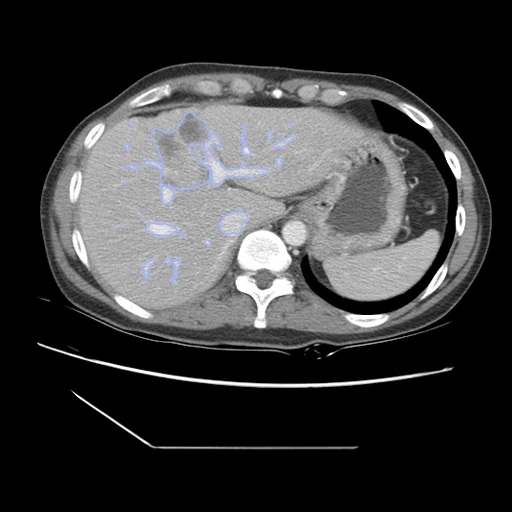
Portal venous CT of the liver of a Memorial Sloan Kettering patient with a primary or metastatic liver tumour, with the liver's vessels and tumours segmented (Medical Segmentation Decathlon, Task08 HepaticVessel).
Read before comparing (8)
- Truth. Label 1 is the liver's vessels: a seed drawn by hand, grown by a level set in the Scout application, then adjusted by hand by an expert abdominal radiologist. Label 2 is the tumours, whose making the data paper does not describe. One reader a scan; the challenge paper says the vessel annotations were found not optimal after the release. The liver is not labelled.
- Vessels. They come in pieces (13 to 92 on the test cases, median 55) and follow the portal vein and the vena cava out of the liver: on hepaticvessel_016, 70% of the vessel label lies inside TotalSegmentator's liver, and all but 0.5% inside its liver, portal and splenic vein and inferior vena cava.
- Tumours. 6.7 to 1007 ml on the test cases, hepaticvessel_209 a mass filling the right lobe, in one piece on every test case but hepaticvessel_057 (two). On hepaticvessel_016, 83% of the tumour lies inside TotalSegmentator's liver and 99% within about 5 voxels of it.
- Images. The decathlon's NIfTI files as delivered: Hounsfield units, the values below -1024 set to -1024, the voxels transposed without resampling to the closest RAS orientation.
- Left out. hepaticvessel_263 to hepaticvessel_460 are patients of TCIA's Colorectal-Liver-Metastases, also Memorial Sloan Kettering's, from which CRLMCT draws its pairs: hepaticvessel_N is CRLM-CT-(N+738). Leaving the range out keeps the two datasets apart by patient.
- Overlap. 181 of the 190 scans in that range share their CRLM scan's slice count and pixel size, the twin of each of CRLMCT's 40 scans among them, and a slice of hepaticvessel_284 equals one of CRLM-CT-1022. The nine training scans outside the range with such a match hold no slice of it; no case built matches a corpus image by slice count and pixel size.
- Room. On the challenge's hidden test set the 19 entries' mean Dice ran from 0.14 to 0.63 on the vessels (median 0.55; nnU-Net 0.63) and from 0.11 to 0.69 on the tumours (median 0.38; nnU-Net 0.69). The surface Dice is at 3 mm, the error the challenge's clinicians would accept.
- Outside numbers. None compare: the challenge ranks its entries on its hidden test set. The test cases here are training cases, public with their labels, so a network trained on the decathlon's task has seen them.
No method has a result on every case of this dataset yet.
Case by case
Click a score to look at that result: the image, the truth in green and the method's map in red. Underlined scores have their image; for the others you see the case without the result.
About the data
- Test cases
- 17 test cases drawn with seed 2026 among the 163 training cases outside development, and 16 development cases, from a pool of 179. The 124 scans of Colorectal-Liver-Metastases are left out (prepare.py), and the labels of the decathlon's 140 test scans were never released.
- Truth
- contours by a program, verified by people the decathlon's two labels: 1 the vessels of the liver, seeded by hand and grown by a level set in the Scout application, then adjusted by hand by an expert abdominal radiologist; 2 the liver's tumours, whose making the data paper does not describe
- Source
- medicaldecathlon.com
arxiv.org/abs/1902.09063
doi.org/10.1038/s41467-022-30695-9
msd-for-monai.s3-us-west-2.amazonaws.com/Task08_HepaticVessel.tar
www.cancerimagingarchive.net/collection/colorectal-liver-metastases
doi.org/10.1038/s41597-024-02981-2 - Licence
- CC BY-SA 4.0
16 cases for tuning, never tested
hepaticvessel_173, hepaticvessel_031, hepaticvessel_181, hepaticvessel_050, hepaticvessel_200, hepaticvessel_042, hepaticvessel_217, hepaticvessel_234, hepaticvessel_197, hepaticvessel_070, hepaticvessel_065, hepaticvessel_255, hepaticvessel_010, hepaticvessel_227, hepaticvessel_208, hepaticvessel_185